39 muscles of lower limb diagram
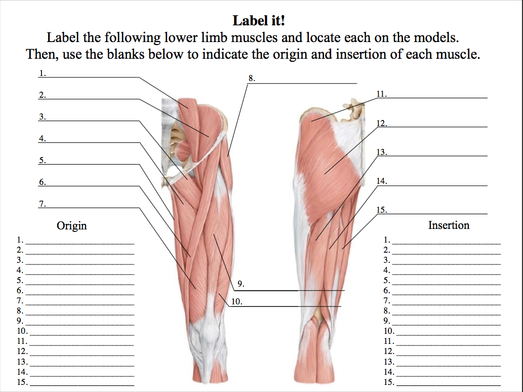
Quizzes on the muscles of the lower limb. The quizzes below each include 15 multiple-choice identification questions related to the muscles of the lower limbs, and includes the following muscles : Adductor longus, Adductor magnus, Extensor digitorum longus, Fibularis brevis, Fibularis longus, Flexor digitorum longus, Biceps femoris, Fibularis ... Lower Leg Muscle Diagram Blank Sketch Coloring Page. Lower Leg Muscle Diagram Blank. India Williams-Shelby. 20 followers . Leg Muscles Anatomy ... The Upper Limb Muscles. The muscles chiefly concerned in producing movements of the joints of the upper limb are as follows : Shoulder. Flexion : pectoralis major, anterior fibres of ...
Start studying Muscles of lower limb (PAL). Learn vocabulary, terms, and more with flashcards, games, and other study tools.
Muscles of lower limb diagram
Plexuses of the Lower Limb "Lumbosacral plexus" Lumbar Plexus Arises from L1-L4 Lies within the psoas major muscle Mostly anterior structures Sacral Plexus Arises from spinal nerve L4-S4 Lies caudal to the lumbar plexus Mostly posterior structures Muscles of the Lower Limb - Listed Alphabetically; Muscle Origin Insertion Action Innervation Artery Notes Image; abductor digiti minimi (foot) medial and lateral sides of the tuberosity of the calcaneus: lateral side of the base of the proximal phalanx of the 5th digit: The lower leg is a major anatomical part of the skeletal system. Together with the upper leg, it forms the lower extremity. It lies between the knee and the ankle, while the upper leg lies between ...
Muscles of lower limb diagram. Lower Limb 5 (Continued) 297 LWBK788-Ch5_297-437_Moorecraft Edition 1 24/01/11 9:24 PM Page 297. 298 PART 2INDIVIDUAL MUSCLES BY BODY REGION OVERVIEW OF THE REGION The lower limb is designed for weight-bearing, balance, and mobility. The bones and muscles of the lower limb are larger and stronger than those of the upper limb, which is necessary ... Beside that, we also come with more related ideas as follows free printable human anatomy coloring pages, lower leg muscle diagram blank and lower limb bones unlabeled. Our goal is that these Leg Anatomy Worksheets pictures gallery can be a direction for you, bring you more references and also make you have a great day. If you're looking for a speedy way to learn muscle anatomy, look no further than our anatomy crash courses. Let's take a look at how you can use muscle diagrams for maximum benefit. Labeled diagram. View the muscles of the upper and lower extremity in the diagrams below. The Lower Limb; Muscles of the Lower Limb; The Fascia Lata. View Article. Muscles of the Gluteal Region. View Article. Muscles of the Thigh. 3 Topics. Muscles of the Leg. 3 Topics. Muscles of the Foot. View Article. Anatomy Video Lectures. START NOW FOR FREE. TeachMe Anatomy. Part of the TeachMe Series.
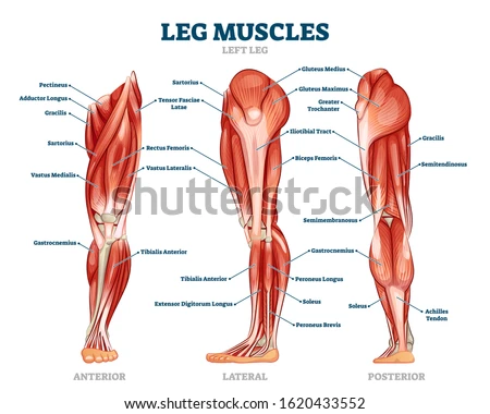
Animal Cell - Color Code the Organelles. Animal Cell Coloring Page. Arteries of the Head and Neck Coloring Page. Arteries of the Lower Limb (Pelvis, Leg and Foot) Coloring Page. Arteries of the Upper Limb (Shoulder, Arm, Hand) Coloring Page. Blood Vessel Anatomy Coloring Page. Blood Vessels (Advanced) Coloring Page. Leg muscles labeled. Take a look at the leg muscles diagram below, where you see each muscle clearly labeled. Spend some time revising this diagram by connecting the name and location of each structure with what you've just learned in the video. The aim of this exercise is to improve your confidence in identifying different structures. Muscles of the Lower Limb Iliacus (part of iliopsoas) ORIGIN: Iliac fossa (ilium); crest of os coxa; ala (sacrum) INSERTION: lesser trochanter (femur) INNERVATION: femoral nerve ACTION: flexes thigh (Anterior view) Muscles Moving Thigh - Anterior Psoas major (part of iliopsoas) ORIGIN: T 12 - (ilium)L 5 Start studying Muscles of the Lower Limb (Posterior View). Learn vocabulary, terms, and more with flashcards, games, and other study tools.
Start studying Muscles of the hip and lower limb. Learn vocabulary, terms, and more with flashcards, games, and other study tools. The arrangement of muscles in the lower limb is sim-ilar to that of the upper limb (see Figure 4.34 and Table 4.14). It must be remem-bered that flexion at the knee results in moving the lower leg posteriorly, unlike the upper limb where flexion of the elbow results in anterior movement of the forearm. The pectineus and iliopsoas muscles are responsible for movement at the hip and are discussed elsewhere. Sartorius: The sartorius, a thin muscle in the thigh, the is the body's longest muscle. Attachments: Originates from the pelvis and attaches to the tibia. Actions: Flexing of the lower leg at the knee joint. The myology of the lower limb is also particularly well represented in this atlas of anatomy, with multiple anatomical charts and diagrams: The first diagram summarizes the different muscular compartments (fascial compartments) of the thigh and leg, and the different fascias (crural fascia, intermuscular septum, interosseous membrane, adductor canal, fascia lata)
Composed of handy tables and diagrams listing attachments, innervation and functions for every muscle, our lower limb muscle anatomy chart will cut your study time in half Mnemonics In order to remember the muscles of the lateral compartment of the leg and their innervation, you can use the following mnemonic:
The muscles of the upper limb can be divided into 6 different regions: pectoral, shoulder, upper arm, anterior forearm, posterior forearm, and the hand.. There are 4 muscles of the pectoral region: pectoralis major, pectoralis minor, serratus anterior and subclavius.Collectively, these muscles are involved in movement and stabilisation of the scapula, as well as movements of the upper limb.
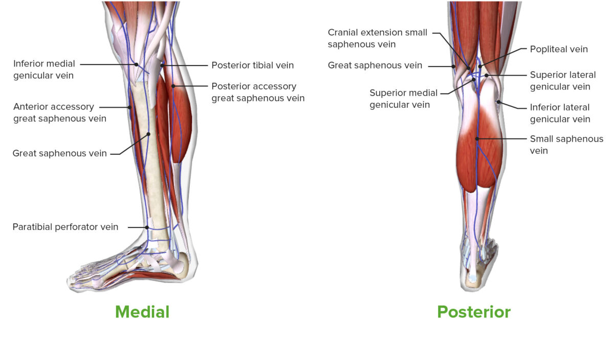
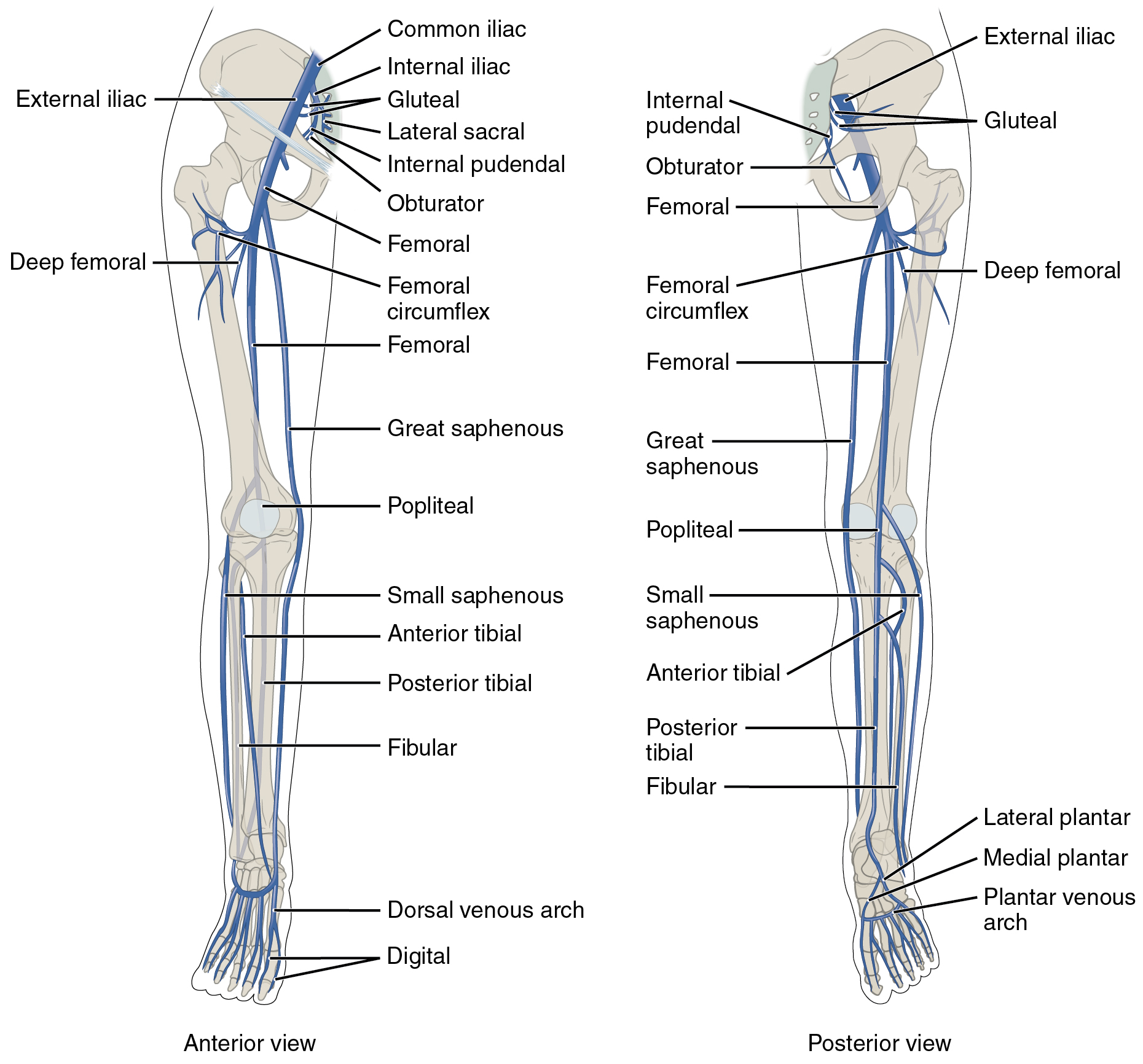
- Bones, Muscles and Ligaments of the Foot - Blood and Venous Supply of the Lower limb - Blood and Venous Supply of the Lower limb - Bones, Ligaments and Muscles of the Spine - Pelvic Floor, Abdominal contents and Muscles, Thoracolumbar fascia These coloured diagrams have been illustrated using a variety of current and reputable sources.
3D interactive models and tutorials on the anatomy of the lower limb, including the muscular compartments, osseus structures, blood supply and innervation.
Key facts about the lower extremity; Hip and pelvis: Bones: hip bones, saccrum, coccyx Hip joint: ball and socket joint Muscles: anterior and posterior (superficial, deep) groups Arteries: gluteal and femoral arteries Veins: external and internali iliac veins Nerves: cluneal, femoral cutaneous, femoral, obturator, sciatic and gluteal nerves, all branches of the lumbosacral plexus
Start studying Muscles of the lower limb. Learn vocabulary, terms, and more with flashcards, games, and other study tools.
Official Ninja Nerd Website: https://ninjanerd.orgNinja Nerds!In this lecture Professor Zach Murphy will present on the anatomy of the leg muscles while usin...
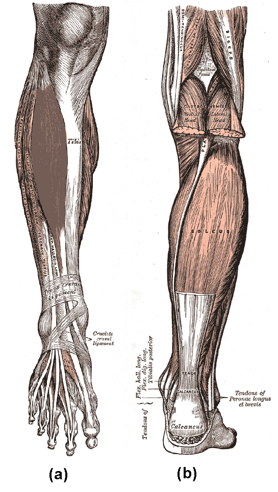
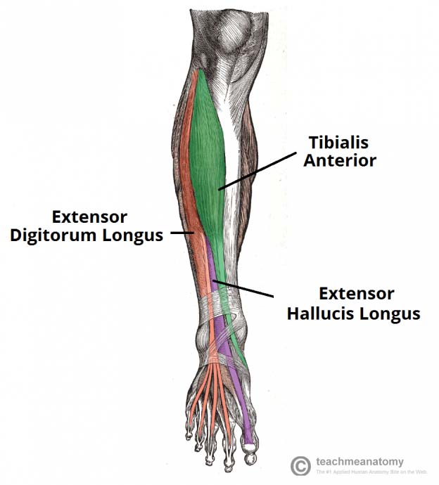
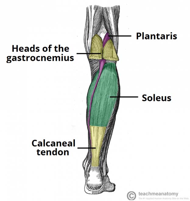
Module 9: Muscles of the Limbs. Search for: Muscles of the lower leg and foot. Information. The muscles of the lower leg, called simply the leg by anatomists, largely move the foot and toes. The major muscles of the lower leg, other than the gastrocnemius which is cut away, are shown in Figure 9-12.
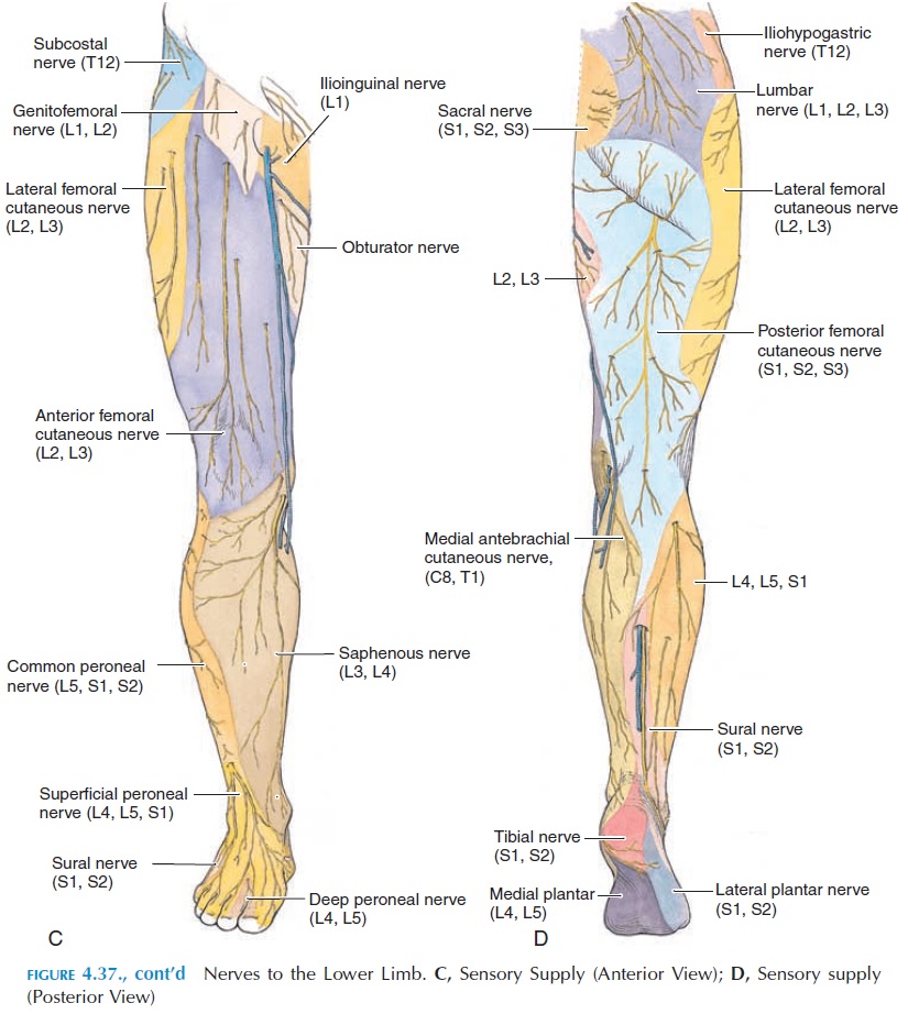
The sciatic nerve is the longest single and continuous nerve in the entire body. It originates from the sacral plexus (L4-S3) and travels all the way down the posterior aspect of the lower limb., The sciatic nerve innervates the entire skin of the leg, the posterior thigh muscles, and the muscles of the leg and foot.
Lower limb (free PDF download) This muscle chart eBook covers the following regions: Inner hip & gluteal muscles. Anterior, medical and posterior thigh muscles. Anterior, lateral and posterior leg muscles. Dorsal and plantar foot muscles. This eBook contains high-quality illustrations and validated information about each muscle.
♦ Thus most of the muscles of the lower limb are supplied by sciatic nerve except the adductors of the thigh and extensors of the knee joint. ♦ Arterial occlusive disease of the lower limb: Occlusive disease causes ischemia of the muscles of the lower limb leading to cramp-like pain. The pain disappears with rest but comes back with activity.

Drawing To Show The Action Of The Calf Muscle In Pumping Blood From The Lower Limb Back To The Heart Royalty Free Cliparts Vectors And Stock Illustration Image 14192059
The lower leg is a major anatomical part of the skeletal system. Together with the upper leg, it forms the lower extremity. It lies between the knee and the ankle, while the upper leg lies between ...
Muscles of the Lower Limb - Listed Alphabetically; Muscle Origin Insertion Action Innervation Artery Notes Image; abductor digiti minimi (foot) medial and lateral sides of the tuberosity of the calcaneus: lateral side of the base of the proximal phalanx of the 5th digit:
Plexuses of the Lower Limb "Lumbosacral plexus" Lumbar Plexus Arises from L1-L4 Lies within the psoas major muscle Mostly anterior structures Sacral Plexus Arises from spinal nerve L4-S4 Lies caudal to the lumbar plexus Mostly posterior structures
:background_color(FFFFFF):format(jpeg)/images/library/10967/unlabelled_muscles_diagram.png)
:background_color(FFFFFF):format(jpeg)/images/library/11153/muscles-tibia-fibula_english__2_.jpg)






:background_color(FFFFFF):format(jpeg)/images/article/en/muscle-anatomy-charts/2mzZyy0wDF3gODSlIc8Okg_sample_page_1.png)






:background_color(FFFFFF):format(jpeg)/images/library/11154/muscles-foot_english__1_.jpg)

0 Response to "39 muscles of lower limb diagram"
Post a Comment