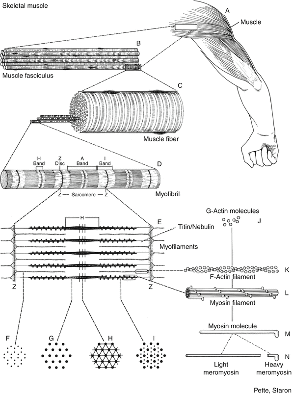
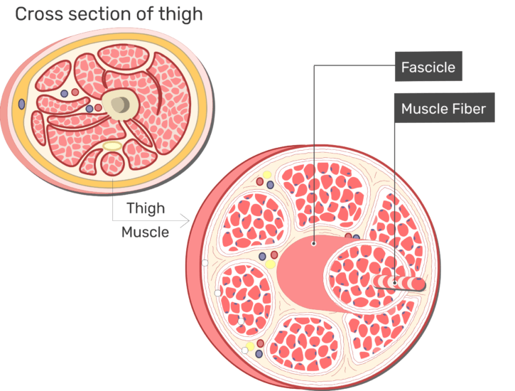
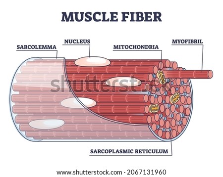
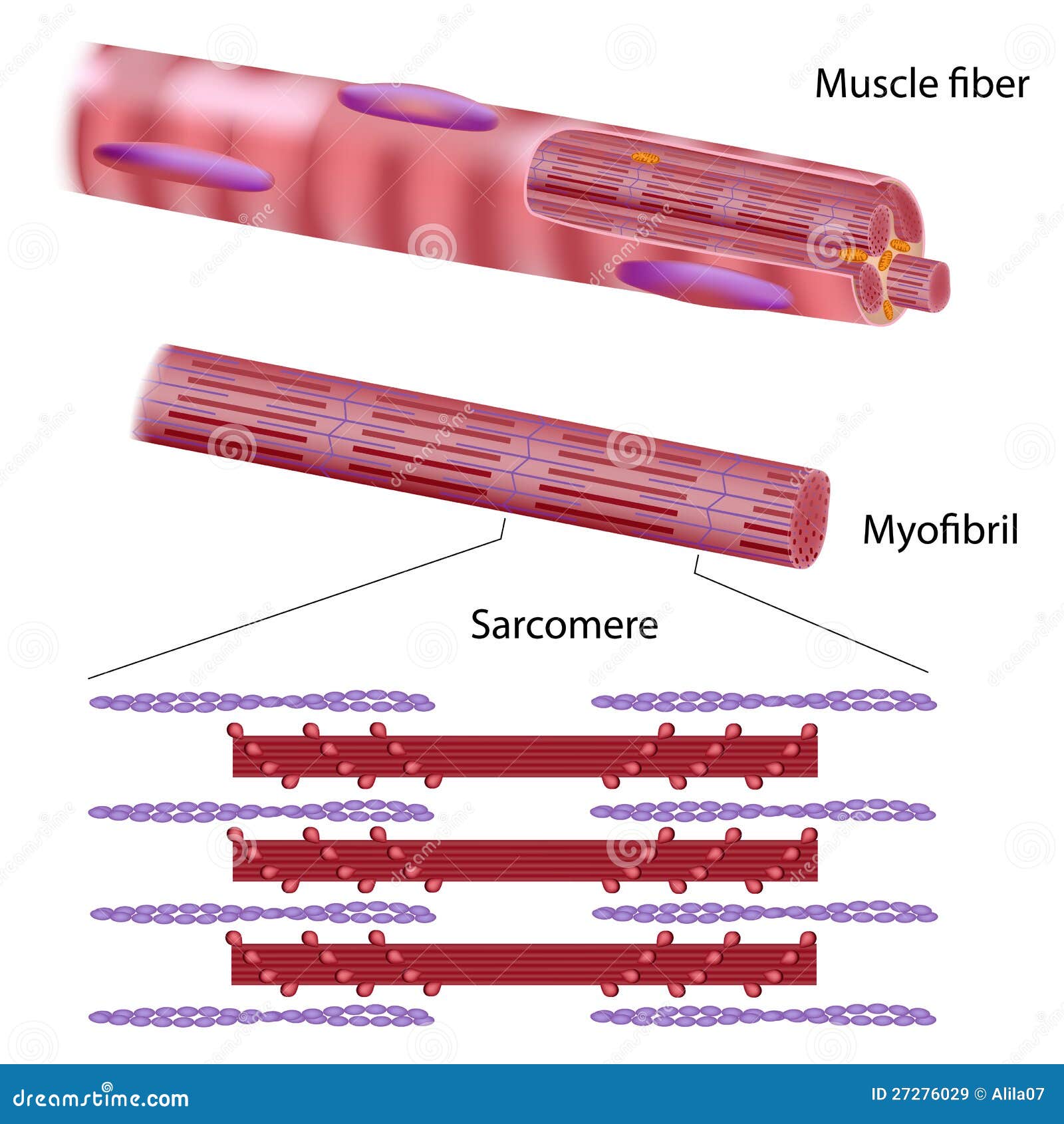
39 diagram of muscle fiber
Cardiac muscle shows many structural and functional characteristics intermediate between skeletal and smooth muscles. Today, in this short article, I will show you the important histological features from the cardiac muscle histology slide. You will get the basic guide to learn cardiac muscle histology with real slide images and labeled diagrams. Cardiac muscle is an involuntary muscle. The heart is an involuntary muscle that beats whether you are awake or asleep. Cardiac muscle contracts to pump blood through the network of veins. Smooth muscles are involuntary, much like a cardiac muscle. They are present in the gastrointestinal system, blood vessels, and body hair.
Muscle fibers Figure 12.3 Motor end plates at the neuromuscular junction. The neuromuscular junction is the synapse between the nerve fiber and muscle fiber. The motor end plate is the specialized portion of the sarcolemma of a muscle fiber surrounding the terminal end of the axon. (a) An illustration of the neuromuscular junction.
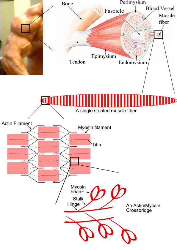
Diagram of muscle fiber
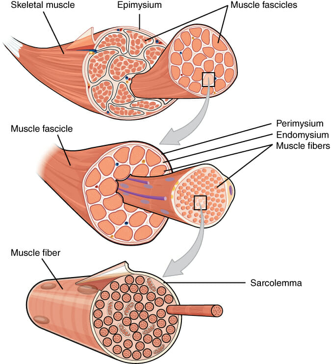
Home » Tanpa Label » Muscle Fiber Anatomy : Skeletal Muscle Anatomy And Physiology I - The membrane of the cell is the sarcolemma; When the nervous system signal reaches the neuromuscular junction a chemical message is . Diagram of a single-fiber electromyography electrode within a motor unit. Light-colored symbols indicate muscle fibers belonging to one motor unit. These muscles allow us to do running, jumping and lifting we do every day. Gross Anatomy of Skeletal Muscle. Each skeletal muscle has three levels of connective tissue. Epimysium is the outer layer of connective tissue surrounding the whole muscle. A bundle of muscle fibers is known as a fascicle.
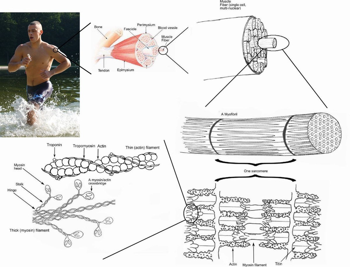
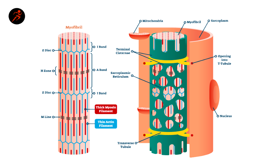
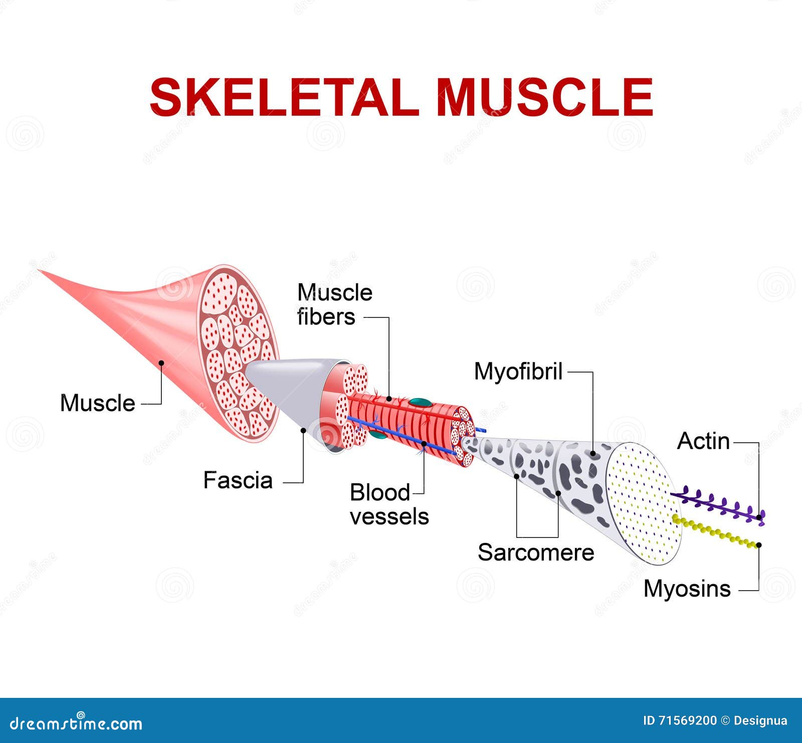
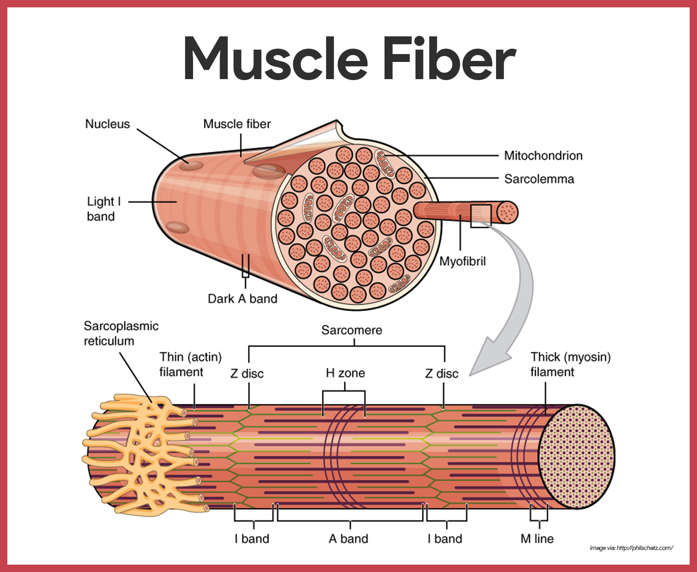
Diagram of muscle fiber. Muscle fibers differ in contraction speed (i.e., slow-twitch, fast-twitch) and whether they primarily generate energy aerobically or anaerobically via the glycogen-lactate system. Structure Of Skeletal Muscle And Motor Units. A. A motor unit consists of a single motor neuron and the muscle fibers innervated by it. B. Epimysium is the same as investing fascia. Actin (thin) and myosin (thick) filaments are contractile elements in the muscle fibers. Whereas the structural unit of a muscle is a skeletal striated muscle fiber ... 12+ Cardiac Muscle Diagram. Cardiac muscle tissue is made up of many interlocking cardiac muscle cells, or fibers, that give the tissue its properties. This is one feature that differentiates it from skeletal. Contraction of Cardiac Muscle - Pathway of Contraction … from teachmephysiology.com Skeletal muscle is composed of cells collectively referred to as muscle fibers. Each muscle fiber is multinucleated with its nuclei located along the periphery of the fiber. Each muscle fiber further subdivides into myofibrils, which are the basic units of the muscle fiber. These myofibrils are surrounded by the muscle cell membrane (sarcolemma ...
Direction Of Muscle Fiber - The Muscular System /. By Drawing Maier on Kamis, 11 November 2021. Muscle contractions occur when two proteins that make up muscle fibers are activated by a nerve to increase the tension within the muscle. Caiaimage / justin pumfrey / getty images muscle contraction occurs when a muscle fiber or group of f. Skeletal muscle is one of the three types of muscles in the human body- the others being visceral and cardiac muscles. In this lesson, skeletal muscles, its definition, structure, properties, functions, and types are explained in an easy and detailed manner. Oblique Muscle Fiber Arrangement : The Muscular System You Got Tickets Naming Muscles -. The anatomical arrangement of skeletal muscle fascicles can be described as parallel, convergent, pennate, or sphincter. Addition to the complex arrangement of muscle fibers,. These are long and thin with fibers running the length of the muscle . Special terms are used to describe structures associated with skeletal muscle tissue. Muscle tissue terms often begin with myo-, mys-, or sarco-. The cytoplasm of a muscle cells is referred to as sarcoplasm.The plasma membrane is called the sarcolemma and the endoplasmic reticulum is called the sarcoplasmic reticulum.A muscle fiber may also be referred to as a myofiber.
Muscles that perform fine movements—like those of the eyes or fingers—have very few muscle fibers in each motor unit to improve the precision of the brain's control over these structures. Muscles that need a lot of strength to perform their function—like leg or arm muscles—have many muscle cells in each motor unit. Blank Muscle Diagram Unlabeled : Muscle Fiber Diagram Unlabeled Png Image Transparent Png Free Download On Seekpng : Posted by Larry Napier on Kamis, 11 November 2021 In this chapter we describe the gross anatomy of the muscular system and consider functional relationships between muscles and bones of the body . Types of neurons and synapse (diagram) Depending on the type of target tissue, there are central and peripheral synapses. Central synapses are between two neurons in the central nervous system, while peripheral synapses occur between a neuron and muscle fiber, peripheral nerve, or gland. Each synapse consists of the: A fasciculus is composed of several muscle cells or muscle fibers. Each muscle fiber is a single cylindrical cell that contains several nuclei located at the periphery of the muscle fiber. The cytoplasm of the muscle fiber contains numerous myofibrils. Each myofibril is a threadlike structure that extends from one end of the muscle fiber to the
As mentioned above, the trapezius muscle is divided into 3 areas: The upper fibers, the middle fibers (called the middle trapezius), and the lower fibers (called the lower traps). The division into the separate, distinct parts of this muscle is about functionality. In other words, each area does something different.
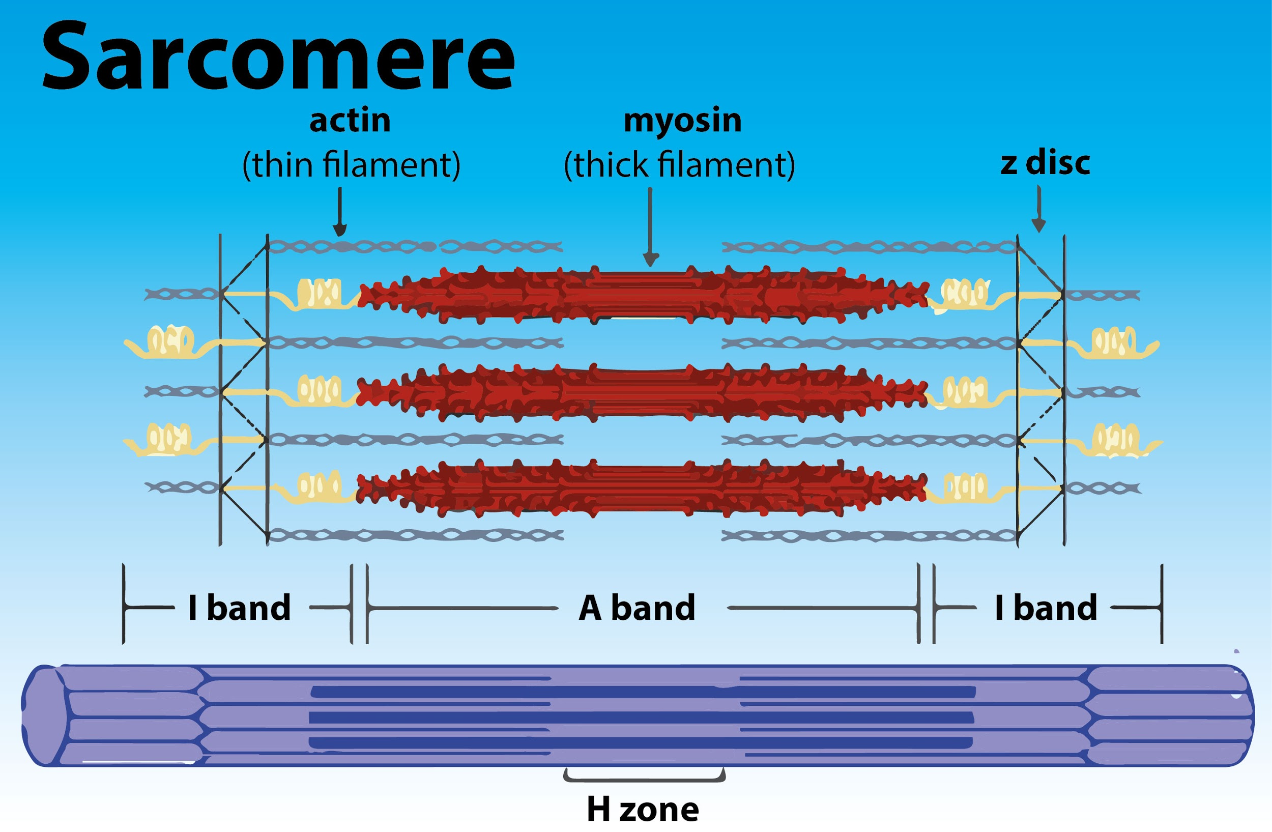
Question 1. Draw the diagram of a sarcomere of skeletal. Solution: Question 2. Define sliding filament theory of muscle contraction. Solution: The mechanism of muscle contraction is best explained by the sliding filament theory which states that contraction of a muscle fiber takes place by sliding of the thin filaments over the thick filaments.
The coordination of the two processes, the transmission of the stimulus and the final contraction of the muscle fiber is called "excitation-contraction coupling". Like nerve fibers, muscle fibers are excitable and are characterized at rest by a so-called resting membrane potential. Various negatively charged particles (anions) and ...
As the diagram shows, most muscles cross an articulation, or joint, to facilitate joint movement. This can be a complex arrangement of bones, muscles, tendons, ligaments, and other connective tissues.
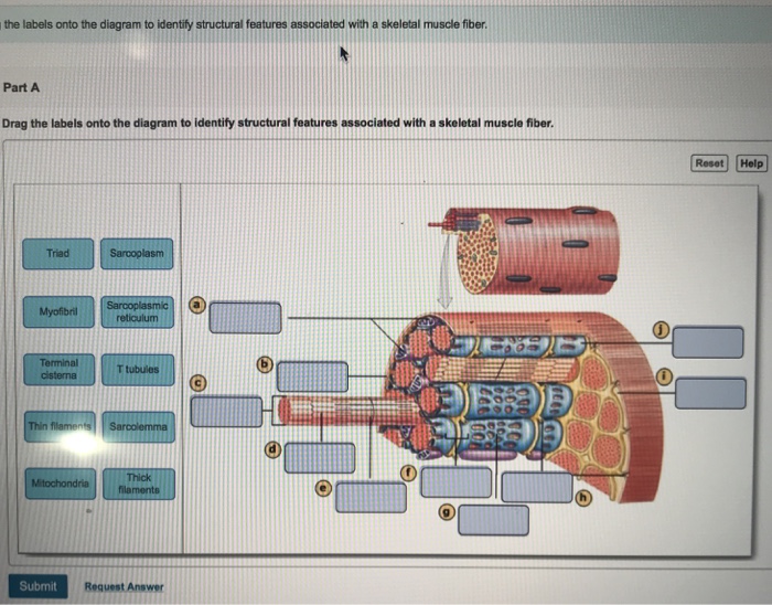
Drag the labels onto the diagram to identify structural features associated with skeletal muscle. An er diagram for a college system is an entity relationship diagram that is used to identify the entities of the college system and what those entities expect from the system. Art-labeling activity structure of a skeletal muscle fiber quizlet ...
Skeletal muscle tissue is composed of long cells called muscle fibers that have a striated appearance. Internal Structure Of Skeletal Muscle Fiber Diagram Quizlet from o.quizlet.com Muscle fibers are organized into bundles supplied by . (b) diagram of part of a muscle fiber . The membrane of the cell is the sarcolemma; The length of a skeletal ...
It is predicted that TNNT3 can regulate muscle growth and muscle fibers 46. TNNT3 is an important part of pig skeletal muscle filaments, which can affect the taste and tenderness of pork 47 , 48 .
This type of muscle creates movement in the body. You have more than 600 muscles in your body! Muscle worksheet with an unlabeled diagram. Human anatomy · human muscular system · human body anatomy · skeletal muscle · muscle fiber · muscle diagram · human skeleton · human muscle structure.
The skeletal muscle fibers are elongated, cylindrical and multinucleated cells whose length may vary in different animals. In this short guide, you will get a basic concept of skeletal muscle histology from the real slide and labeled diagram. You will also get the identification points of skeletal muscle histology slide with a little description here in this guide.
The masseter muscle fibers converge inferiorly, forming a tendon that inserts the outer surface of the mandibular ramus and coronoid process of the mandible. The function of the masseter muscle is to elevate the mandible and approximate the teeth—additionally, the intermediate and deep muscle fibers of the masseter function to retract the ...
The humidity-responsive cotton artificial muscle fibers were constructed from twisted and plied cotton fibers, which used torque-balanced fiber structures to avoid the need for torsional tethering. The cotton artificial muscle fiber produced a completely reversible torsional stroke of 42.55° mm −1 , which was at the same level as that of the ...
These muscles allow us to do running, jumping and lifting we do every day. Gross Anatomy of Skeletal Muscle. Each skeletal muscle has three levels of connective tissue. Epimysium is the outer layer of connective tissue surrounding the whole muscle. A bundle of muscle fibers is known as a fascicle.
Diagram of a single-fiber electromyography electrode within a motor unit. Light-colored symbols indicate muscle fibers belonging to one motor unit.
Home » Tanpa Label » Muscle Fiber Anatomy : Skeletal Muscle Anatomy And Physiology I - The membrane of the cell is the sarcolemma; When the nervous system signal reaches the neuromuscular junction a chemical message is .







:watermark(/images/watermark_only.png,0,0,0):watermark(/images/logo_url.png,-10,-10,0):format(jpeg)/images/anatomy_term/nucleus-of-skeletal-muscle-fiber/3fBKxdMoyUFj8fsymsuRrw_134Nucleus_of_skeletal_muscle_fiber_magnified.png)




0 Response to "39 diagram of muscle fiber"
Post a Comment