39 which part of the diagram is considered nerve fiber
Principles of Anatomy and Physiologych 12 - Prometheus. Course: StuDocu Summary Library EN. Tortora and Derrickson Principles of Anatomy & Physiology, 12 th edition. Ch12: Nervous Tissue. Multiple Choice. 1.
1) Which part of the diagram is considered nerve fiber? a) A b) D c) I d) Both A and D e) All of these choices make up the nerve fiber
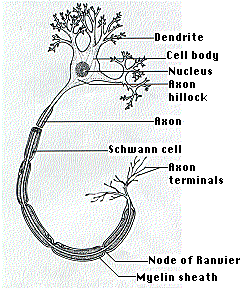
The unmyelinated parts of the nerve fiber are nodes of Ranvier. This way of action potential propagation is called saltatory conduction (red arrows in the diagram) Ion channels open, allow sodium ions to enter the cell leading to membrane depolarization and generation of action potential.
Which part of the diagram is considered nerve fiber
The root of the penis is the most proximal part of the penis. It is located in the urogenital triangle of the perineum, where it is fixed to the pubic symphysis via the two suspensory ligaments of the penis.The root consists of the two muscles (ischiocavernosus and bulbospongiosus muscles) and proximal expansions of the erectile tissues; the two crura of penis and the bulb of penis.
Nerve fiber refers to: a. Axon. b. Dendrites. c. Nissl body. d. Both a and b. e. All of the above. D. 7 ... Sodium pumps are considered electrogenic because. a. They contribute to the negativity of the resting membrane potential ... Which part of the diagram is considered nerve fiber? a. A b. D c. H d. Bothaandb e. All of the above. E. Decks in ...
6. Ganglion cell fiber layer. The ganglion cell axons run in the nerve fiber layer above the inner limiting membrane towards the optic nerve head in a arcuate form (Fig. 00, streaming pink fibers). The fovea is, of course, free of a nerve fiber layer as the inner retina and ganglion cells are pushed away to the foveal slope.
Which part of the diagram is considered nerve fiber.
Which part of the diagram is considered nerve fiber? a) A b) D c) I d) Both A and D e) All of these choices make up the nerve fiber. D. This part of the neuron contains the nucleus and Nissl bodies. a) A b) B c) C d) E e) Both A and B. B.
Which part of the diagram is considered nerve fiber? A, B. This part of the diagram contains organelles and Nissl bodies. E. This portion of the diagram contains cytoplasm and a myelin sheath wrapped around neurolemma. C. In the diagram, where is the axon collateral? H.
Which part of the diagram is considered nerve fiber? A D I Both A and D All of these choices make up the nerve fiber Both A and D Na+/K+-ATPase is considered to be an electrogenic pump because A. it contributes to the negativity of the resting membrane potential. B. the sodium ions are negatively charged. C. it exhibits low permeability.
Which part of the diagram is considered nerve fiber? a) A b) D c) I d) Both A and D e) All of these choices make up the nerve fiber. d. In the diagram, where are axon terminals? a) F b) G c) H d) I e) None of these choices. c) H. This part of the neurons contains the nissl bodies. a) A b) B c) C d) D e) both A and B. B.
Which part of the diagram is considered nerve fiber? A)A B)D C)I D)Both A and D E)All of these choices make up the nerve fiber. Free. Multiple Choice . Unlock to view answer. Q 37 Q 37. This part of the neuron contains the nucleus and Nissl bodies. A)A B)B C)C D)E E)Both A and B. Free.
Which part of the diagram is considered nerve fiber? all of these choices make up the nerve fiber both a and d a I d. c. remove a neurotrasmitter. Diffusion, enzymatic degradation, and uptake by cells are all ways to a. inhibit a presynaptic potential b. stop a spatial summation
Section Reference 1: Sec 12.2 Histology of Nervous Tissue. 36) This structure electrically insulates the axon of a neuron to increase the speed of nerve impulse conduction. a) A b) Bc) C d) D e) E Answer: e. Difficulty: EasyStudy Objective 1: SO 12.2 Compare the structures and functions of neurons and neuroglia and white matter and gray matter.
Phone: 602-282-1547; info@bestjanitorialservices.com; Home; About Us; Services. Commercial Cleaning Services; Construction Cleaning Services
Which cranial nerve in the diagram is primarily involved in the sense of vision? B. 57. ... Which is NOT considered an accessory structure of the eye? Retina. 66. ... Which part of the diagram contains the olfactory bulb neurons? A. 74.
69) The function of this pathway is to convey nerve impulses from the brainstem to cause automatic movements that regulate muscle tone, posture, and balance and orientation of the head and body. a) Indirect pathway
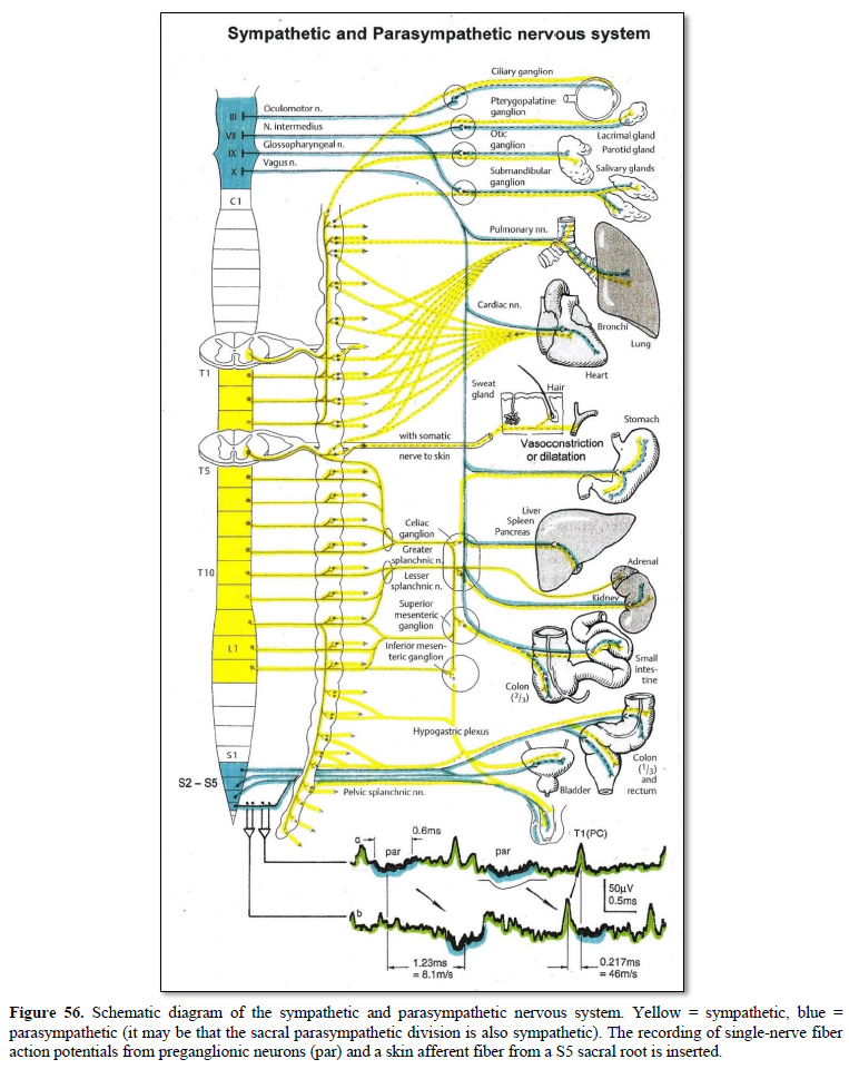
To continue with the analogy of the circuit diagram, there are three different types of "junctions" that operate within the sympathetic system ().The first type is most direct: the sympathetic nerve projects to the chain ganglion at the same level as the target effector (the organ, tissue, or gland to be innervated). An example of this type is spinal nerve T1 that synapses with the T1 ...
Saphenous Nerve It is the largest cutaneous branch of the femoral nerve. It supplies cutaneous branches to the skin of the leg and foot in the region between the knee and the ankle. Sciatic Nerve Also known as the ischiatic nerve, the sciatic nerve is a nerve fiber that begins in the lower back and ends in the lower limb.
Scitech - classification and identification of human ...
Taste buds are microscopic sensory organs containing chemosensory cells which synapse with afferent fibers of gustatory nerves. The number of taste buds in the oral cavity and uppermost gastrointestinal tract is subject to a high degree of interindividual variation (500-5000) while the number of cells in one taste bud can be up to 150 Due to the abrasive environment of the oral cavity ...
.jpg)
Peripheral nerve fiber types | mountain west foot & ankle ...
Mixed cranial nerves are the cranial nerves that contain sensory and motor nerve fibers. There are four of such nerves in our peripheral nervous system ; Trigeminal nerve (CN V) Facial nerve (CN VII) Glossopharyngeal nerve (CN IX) Vagus nerve (CN X)

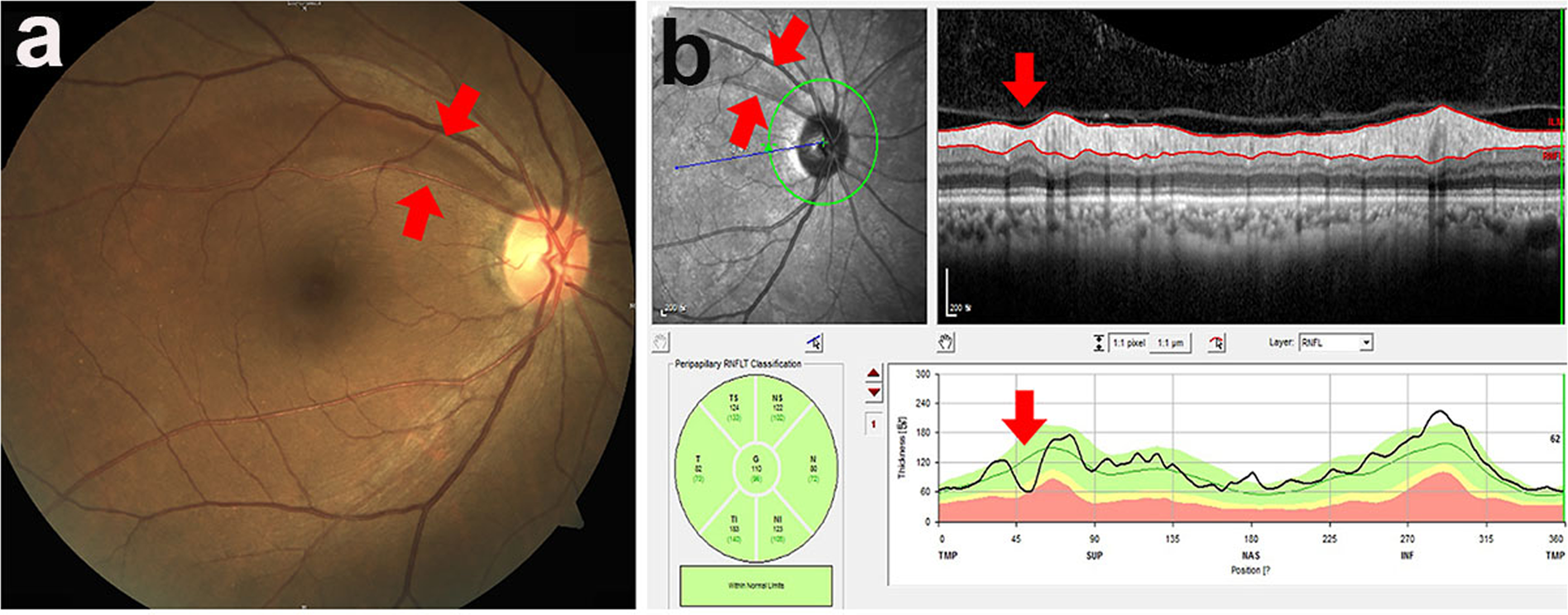
Sex-specific differences in circumpapillary retinal nerve ...
Whereas axons originating from lmns are considered part of the cranial nerves themselves. Which part of the diagram is considered nerve fiber. This part of the neuron contains the nucleu and nissl bodies. Which part of the diagram is considered nerve fibera ab dc id both a and de all of these choices make up the nerve fiber.

Electrical diagram of the new sensitive nerve fiber model ...
Use The Diagram To Determine What Flower Structures Develop Under The Conditions Described Below. The structure of plant cells can vary during the early stages of growth. In the chart below identify the structures that will de…. Written By Maria M Beus Friday, December 22, 2017 Add Comment.

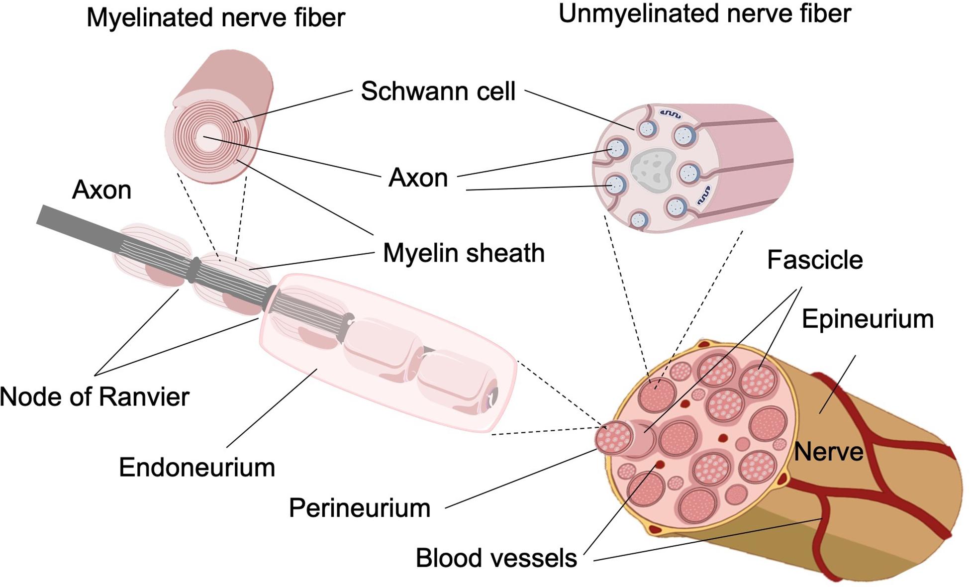
A nerve is a cordlike organ composed of numerous nerve fibers ...
Which part of the diagram is considered nerve fiber? Question options: D choices make up the nerve fiber. Which of the diagrams in the figure represents a ligand-gated channel? Question options: 1 / 1 point. Share this link with a friend: Copied! Other Related Materials.

Ijms | free full-text | bioactive nanofiber-based conduits in ...
Unlike other nerves of the body that are myelinated by Schwann cells, the fibers of the optic nerve are myelinated by oligodendrocytes. Arising from the posterior pole of each eye, each optic nerve is about 35 mm and 55 mm in length and can be subdivided as follows: The optic nerve head. The intraorbital part. The intracanalicular part.
Ijms | free full-text | a novel in vitro assay using human ...

Time-course changes in optic nerve head blood flow and ...
Nerve fiber, drawing stock photo - alamy

Schematic of the structure of the nerve fiber and the ...
Mengapa akson sering disebut serabut saraf? - quora

Efferent nerve fiber - an overview | sciencedirect topics
Early corneal nerve fibre damage and increased langerhans ...

Nerve fiber types - sciencedirect
Association between localised retinal nerve fibre layer ...

Optical coherence tomography structural abnormality detection ...

Modeling of nerve fibers. (a) schematic drawing of a nerve ...

Wide-field trend-based progression analysis of combined ...

Chapter 12 diagrams from test bank flashcards | quizlet

Which part of the diagram is considered nerve fiber a a b d c ...

Exposure to secondhand smoke in children is associated with a ...
Apa itu serat saraf? - quora

Figure, unmyelinated nerve fibers, myelin sheath ...
Neurons

A) schematic of the nerve fiber with paranode, (b) the ...

Impact of artifacts from optical coherence tomography retinal ...

Chapter 12 diagrams from test bank flashcards | quizlet
Cureus | bilateral peripapillary retinal nerve fiber layer ...

Chapter 12 diagrams from test bank flashcards | quizlet

Nerve cell

Ethnicity-specific database improves the diagnostic ability ...

Neuro clickers 1 flashcards | quizlet

Optical coherence tomography normative peripapillary retinal ...
Frontiers | dendritic degeneration of human auditory nerve ...

A simplified illustration of the general anatomy of the skin ...
Frontiers | biomimetic approaches for separated regeneration ...

File:propagation of action potential along myelinated nerve ...

Optical clearing reveals tnbs-induced morphological changes ...
0 Response to "39 which part of the diagram is considered nerve fiber"
Post a Comment