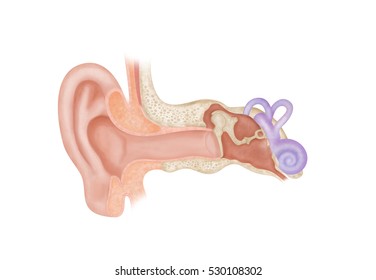
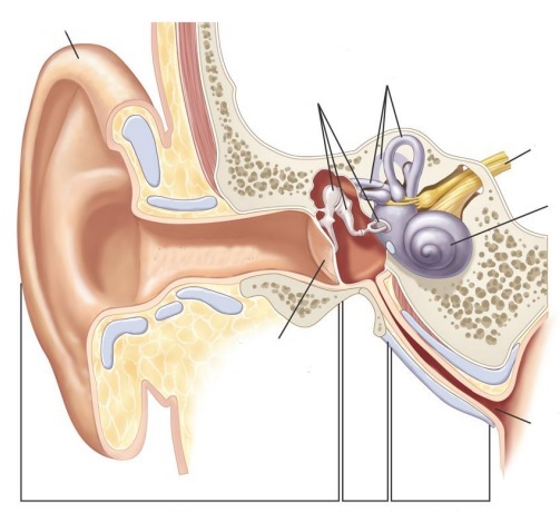
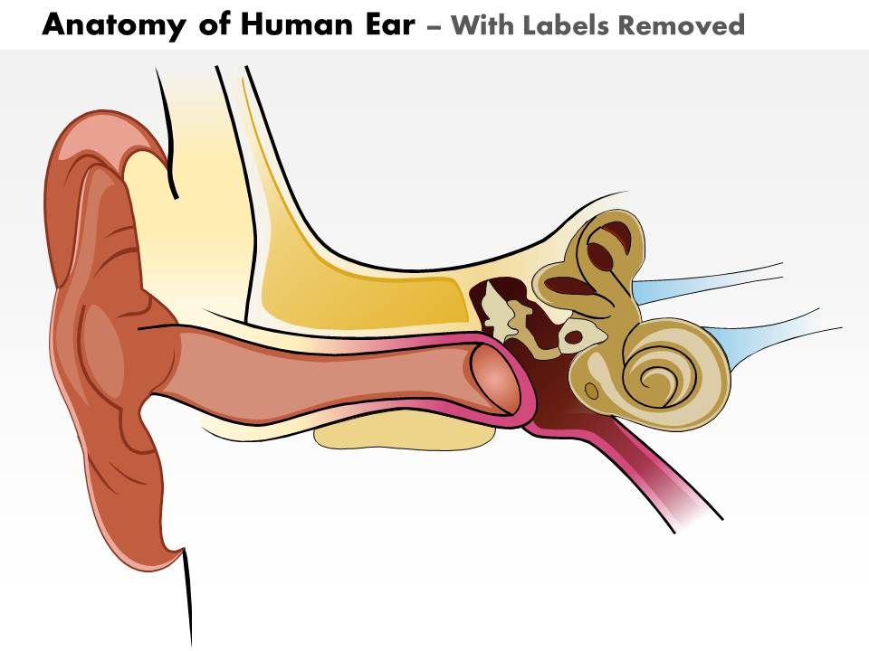
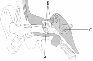
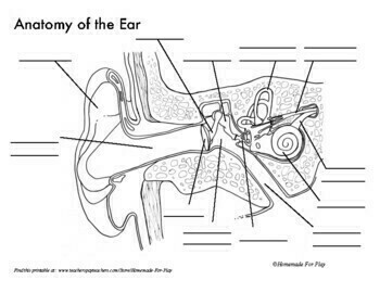
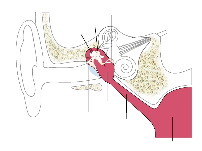
40 ear diagram without labels
Simple Human Ear Diagram Human Ear Diagram Wiring Diagram For You. ... Human Heart Diagram Without Labels Human Heart Diagram Without. Human Heart Drawing Color And Label Basic Diagram Books Charcoal. Simple Diagram Of Lung Wiring Diagram Used. To Draw A Human Heart With Labels Result Unique Simple Diagram Jpg.
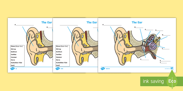
The second page shows an ear diagram without labels. The final page shows the labels linking to the beginning letters of each feature, but without the words list. With this Parts of the Ear Labelling Activity, you can test children's memory and their ability to connect different visual signs with words and letters.
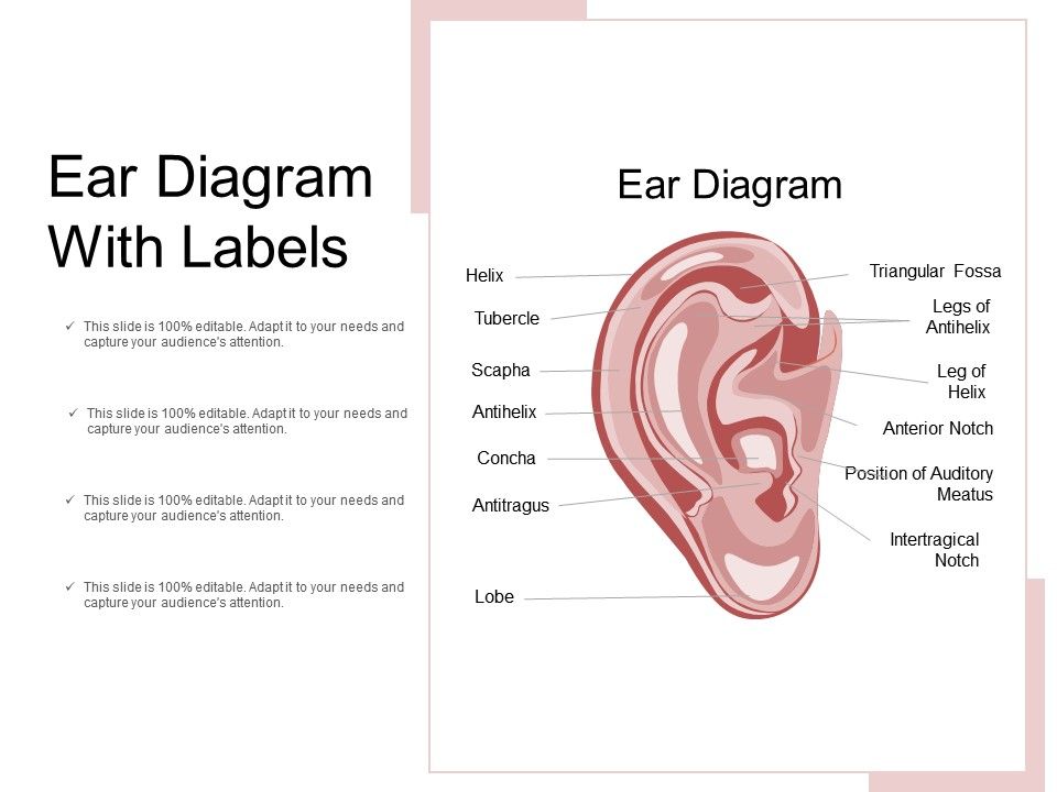
Human Ear Diagram. Wondering what is the structure of the human ear, and how it performs the function of hearing? Look no further, this Bodytomy article gives you a labeled human ear diagram and also explains the functions of its different components.

Ear diagram without labels
Human Heart Diagram Without Labels. Saved by Susan Wells. 5. Carotid Artery Cardiovascular System Tricuspid Valve Human Heart Diagram Physiology Biology Leet Anatomy And Physiology Human.
The sound waves are collected by the external ear up to some extent. They pass through the external auditory meatus to the tympanic membrane which is caused to vibrate. The vibrations are transmitted across the middle ear by the malleus, incus and to the stapes bones. The latter fits into the fenestra ovalis.
How to Draw an Ear - Step-by-Step 1. I started this drawing by blocking in the helix outline. At this early stage, I began to gauge proportions and angles. With the helix blocked in, I marked down where I thought the tragus and anti-tragus envelop the section leading up to the ear canal.
Ear diagram without labels.
12,612 human ear anatomy stock photos, vectors, and illustrations are available royalty-free. See human ear anatomy stock video clips. of 127. the ear anatomy of ear ear anatomy the human ear anatomy of the ear ear diagram ear structure diagram of ear inner middle ear human ear parts. Try these curated collections.
Try this amazing Ear Diagram Quiz quiz which has been attempted 4864 times by avid quiz takers. Also explore over 12 similar quizzes in this category. Take Quizzes. ... Have you been having pain from an ear infection that will come on fast without improving for several days? This may be due to a couple of reasons and one way to self-diagnose ...
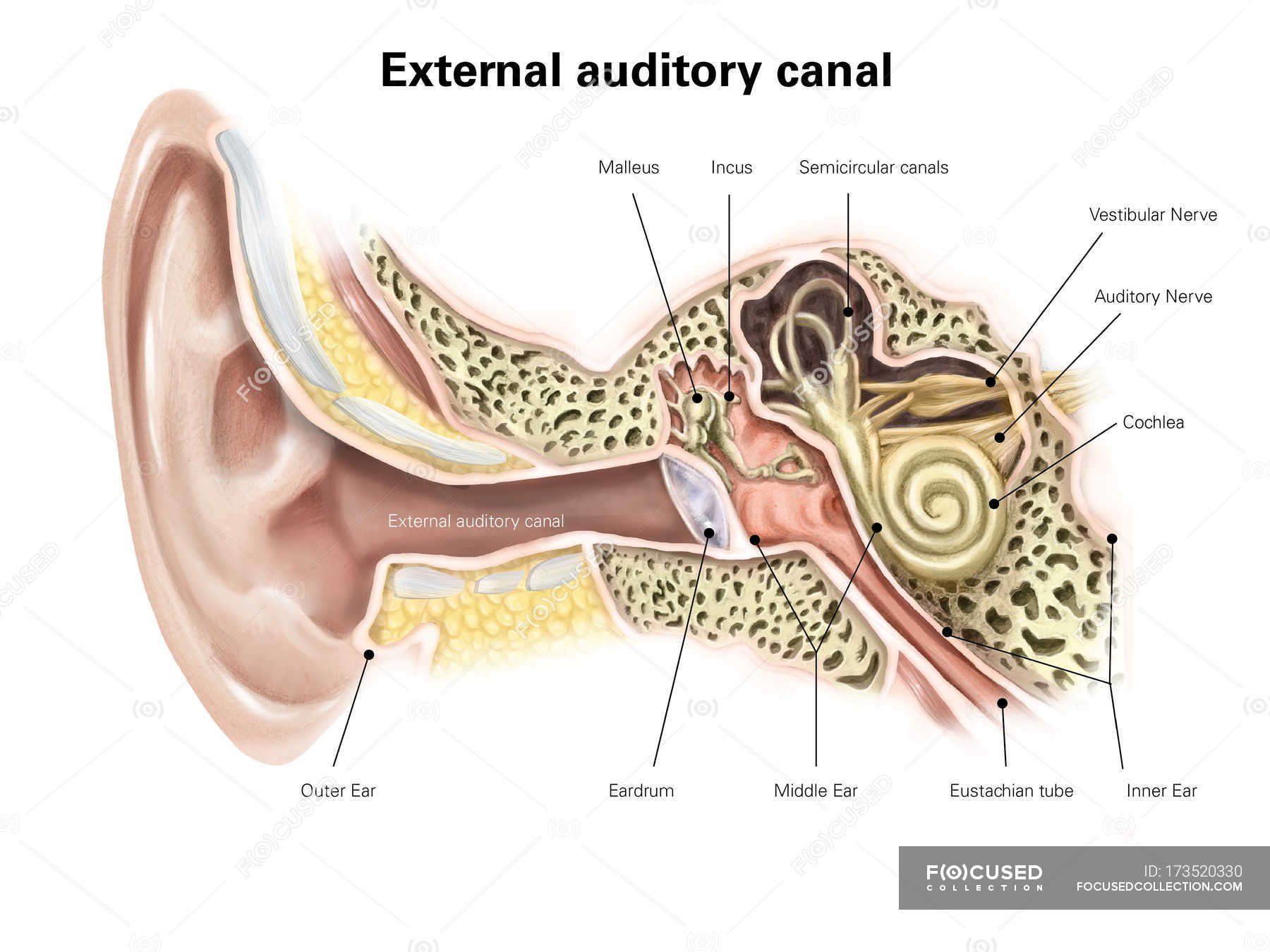
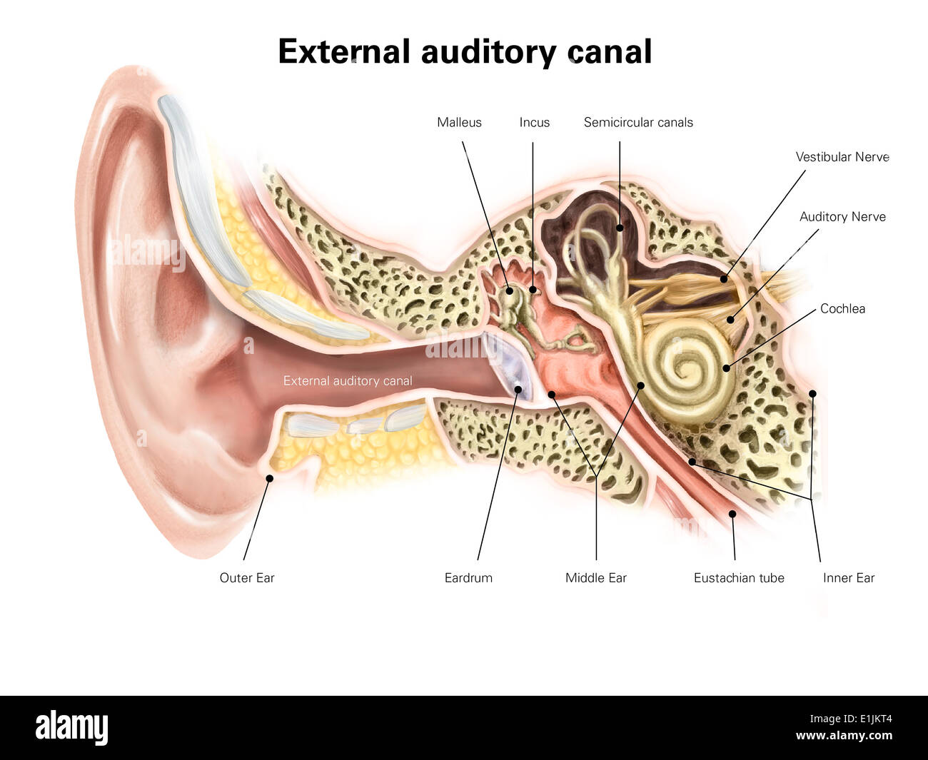
The middle ear is a small, air-filled space containing three tiny bones called the malleus, incus and stapes but collectively called the ossicles. The malleus connects to the eardrum linking it to the outer ear and the stapes (smallest bone in the body) connects to the inner ear. The inner ear has both hearing and balance organs.
A brief description of the human ear along with a well-labelled diagram is given below for reference. Well-Labelled Diagram of Ear The External ear or the outer ear consists of: Pinna/auricle is the outermost section of the ear. The external auditory canal links the exterior ear to the inner or the middle ear.
Take a look at the diagram of the eyeball above. Here you can see all of the main structures in this area. Spend some time reviewing the name and location of each one, then try to label the eye yourself - without peeking! - using the eye diagram (blank) below. Unlabeled diagram of the eye. Click below to download our free unlabeled diagram of ...
Ear Tests. Ear exam: The first test for an ear problem is often just looking at the ear.An otoscope is a device to look into the ear canal to see the drum. Auditory testing: An audiologist ...
A diagram of the ear with Spanish labels. CONTACT US. 825 S. Taylor Avenue Saint Louis, MO 63110. Toll free: 877.444.4574 Tel: 314.977.0132 Fax: 314.977.0023
Ear Anatomy. Detailed Coloring Pages. Anatomy of the Ear (Coloring) Image of the ear is colored according to the directions where structures such as the tympanum, malleus, incus, stapes, and cochlea are indicated. This worksheet is intended to help students learn the parts of the eye. diannedemoss121. D.
The first worksheet presents an ear with annotations showing the first letters of its key features. For example, a label marked 'P' links to the Pinna (outer ear). The second page shows an ear diagram without labels. The final page shows the labels linking to the beginning letters of each feature, but without the words list.
Eye Diagram Handout Author: National Eye Health Education Program of the National Eye Institute, National Institutes of Health Subject: Handout illustrating parts of the eye Keywords: parts of the eye, eye diagram, vitreous gel, iris, cornea, pupil, lens, optic nerve, macula, retina Created Date: 12/16/2011 12:39:09 PM
Spine Diagram without Labels. Eye Diagram without Labels. Spine. Blank Digestive System. Urinary System. Lung. Printable Heart. Skeleton Diagram without Labels. Ear Diagram without Labels.
Structure and Functions of the Ear Explicated With Diagrams. The ear is another extraordinary organ of the house of wonders, that is, the human body. The ear catches sound waves and converts it into impulses, that the brain interprets, making it understandable and helps the human body differentiate between different sounds.
Label Parts of the Human Ear. Select One Auditory Canal Cochlea Cochlear Nerve Eustachian Tube Incus Malleus Oval Window Pinna Round Window Semicircular Canals Stapes Tympanic Membrane Vestibular Nerve. Select One Auditory Canal Cochlea Cochlear Nerve Eustachian Tube Incus Malleus Oval Window Pinna Round Window Semicircular Canals Stapes ...
Parts flower diagram without labels. The stigma is located at the tip of the pistil. Parts flower diagram without labels plant structure and anatomy picture. Cortex function in plants. 11 14 ks3 14 16 ks4 post 16 plant growth health and reproduction. Alternatively your children can label the diagrams to reinforce the topic vocabulary.
Jun 22, 2020 - Heart Diagram Worksheet Blank - 25 Heart Diagram Worksheet Blank , Flower Diagram without Labels - thefrangipanitree
Constricted ear: In this case, the helical rim folds over, is wrinkled, or abnormally tight. Cryptotia: Due to malformation of ear cartilage, this variant gives off the appearance that the upper portion of the ear is buried inside the head. Microtia: This is an underdeveloped ear. Anotia: In some cases, there is a complete absence of the ear.
Ear Anatomy Diagram Printout. EnchantedLearning.com is a user-supported site. As a bonus, site members have access to a banner-ad-free version of the site, with print-friendly pages.
The human body has three layers of skin; Epidermis: It acts as a physical barrier and its outermost layer of the skin. Dermis: It is the middle layer of the skin and consists of sweat glands, hair follicles and connective tissues. Hypodermis: The deepest layer of the ski and is mostly made up of lipids and connective tissues.
Use these simple eye diagrams to help students learn about the human eye. Three differentiated worksheets are included: 1. Write the words using a word bank2. Cut and paste the words3. Write the words without a word bank Labels include: eyebrow, eyelid, eyelashes, pupil, iris, and sclera.UPDATE:I'
Posted on June 7, 2016 by admin. Picture Of Female Reproductive System Diagram 1024×1204 Diagram - Picture Of Female Reproductive System Diagram 1024×1204 Chart - Human anatomy diagrams and charts explained. This diagram depicts Picture Of Female Reproductive System Diagram 1024×1204 with parts and labels.
Anatomy of the Ear. The ear is made up of three parts: the outer, middle, and inner ear. All three parts of the ear are important for detecting sound by working together to move sound from the outer part through the middle and into the inner part of the ear. Ears also help to maintain balance.
Ear Diagram Unlabeled Worksheet asks children to label the parts of the ear and re-arrange a jumbled up Carnivores, omnivores and herbivores Venn diagram. Students knowledge of unlabelled diagram of the human ear diagrams; diagram heart diagrams. Knowledge of biology human lung erica l. Cell wiringall.com - Stačí.
Human ear. The ear is divided into three anatomical regions: the external ear, the middle ear, and the internal ear (Figure 2). The external ear is the visible portion of the ear, and it collects and directs sound waves to the eardrum. The middle ear is a chamber located within the petrous portion of the temporal bone.
Human Ear - Anatomy, Parts (Outer, Middle, Inner), Diagram. The human ear can be divided into 3 parts - external, middle and internal - with each part playing an integral role in the sense of hearing, while the internal ear has an added function for equilibrium. The external (outer) and middle ear transmit sound waves to the internal ...




:background_color(FFFFFF):format(jpeg)/images/library/7459/Ear.png.jpg)









0 Response to "40 ear diagram without labels"
Post a Comment