42 spine l5 s1 diagram
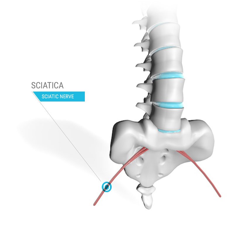
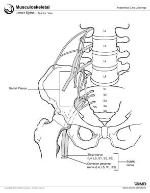
BACKGROUND CONTEXT: Lumbosacral transitional vertebrae (LSTVs) are a congenital vertebral anomaly of the L5-S1 junction in the spine. This alteration may contribute to incorrect identification of a vertebral segment, leading to wrong-level spine surgery and poor correlation with clinical symptoms. The L5 and S1 nerves are near the SI joint and studies have shown that SI joint dysfunction can cause pain and other symptoms in the distribution of these nerves. The SI joint is separate from the sciatic or spinal nerve(s); however, the SI joint can cause sciatica-like symptoms .
The two nerves most commonly pinched in the lower back are L5 (lumbar 5) and S1 (sacral 1). Pinched nerve at L5. The L5 nerve supplies the nerves to the muscles ...

Spine l5 s1 diagram
Lastly is the sacrum, a large triangular bone at the base of the spine. It is formed by the fusing of sacral vertebrae S1-S5. Finding the Conus. You can either count from the sacrococcygeal area there will be 5 sacral and 5 lumbar vertebrae, the conus medullaris should end at of before L2. Furthermore, we attached it along the spine, including S1-L1, L1/T12-C7, C1-C7, and S1-C7 (C, T, L, and S represent the cervical, thoracic, lumbar, and sacrum segments of the spine ... Anatomy of the lumbar spine (CT scan) This anatomy module is dedicated to interns and students that wish to learn more about the anatomy of the lumbar spine in CT. It allows to differentiate the vertebrae, the nervous system, the intervertebral discs and the zygapophyseal joints. The images are available in the three planes, axial, sagittal ...
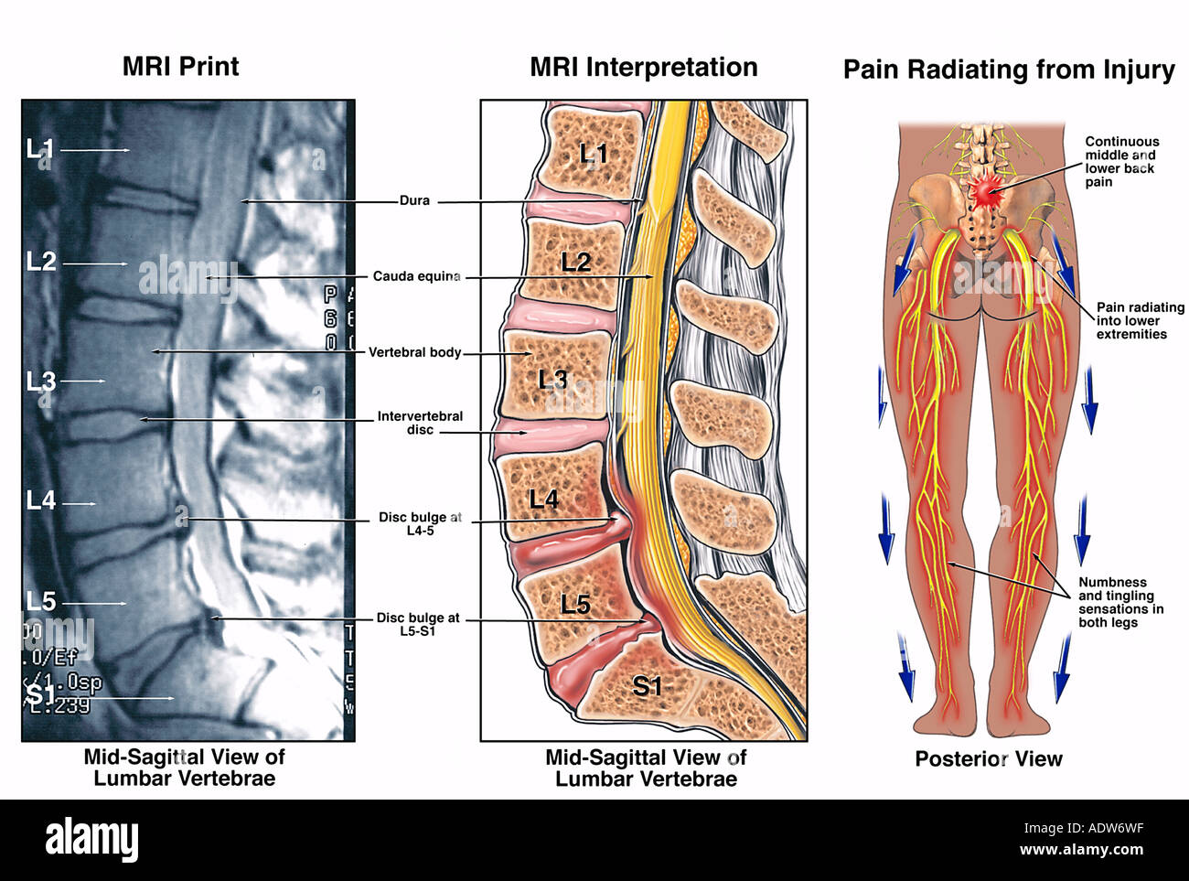
Spine l5 s1 diagram. The relevant anatomy of the spinal nerve-muscular innervation of the back is centered around the lumbar spinal nerves, peripheral nerves of the lumbar plexus, spinal cord, and lumbar vertebral column. Within the lumbar region, the vertebral bodies are larger than in the thoracic and cervical regions due to the lumbar spine being designed for weight-bearing purposes. L5-S1 joint-related problems. As the L5 and S1 vertebrae suffer excess amounts of stress due to their critical location and function, the joint may develop many abnormalities with increased age. Some of these abnormalities are mentioned below: Osteoarthritis: This is the wear and tear type of arthritis. It develops due to aging and because of ... In 1595, French anatomist Andre du Laurens first described the structure of a rope-like tail of fibers at the caudal end of the spinal cord. This bundle of numerous axons was termed the cauda equina, from the Latin translation meaning "horse's tail," and it contains nerves which innervate both sensory and motor targets within lumbar, sacral, and coccygeal spinal cord levels. protrusion专题🌟整理关于protrusion是什么意思protrusion翻译transformational是什么意思protuberance是什么意思ointment💖 ...
Conditions affecting the L5-S1 spinal motion segment are usually treated with nonsurgical methods. If the lower back and/or leg symptoms worsen or do not ... Learn the vertebral column anatomy of the spine or backbone, including the vertebrae or bones of the cervical, thoracic, lumbar, and sacral spine. Vertebrae by definition function to protect the spinal cord and are located around the cord. Learn the anatomy of the spinal column and each vertebra using labeled diagrams and charts. The loose posterior part of the spine is removed, called a Gill laminectomy and the nerve roots of L5 and S1 are inspected and decompressed, as needed. Fixation is then placed at L4, L5 and S1. Since we don't need to move L5 we can place our S1 screws into the L5 body, to give additional fixation strength to the construct. L4-L5 motion segment provides a bony enclosure for the cauda equina which means nerves that continue down from the spinal cord and other fragile structures. Common Injuries take Place at L4-L5 Spine Segment. What makes L4-L5 motion segments prone to facet joint-related issues is the high degree of mobility.
cervical spinal nerves C3 and C4 (motor and sensation) -Actions rotation, retraction, elevation, and depression of scapula. -Antagonist. =serratus anterior muscle, =Latissimus dorsi. levator scapulae. -Origin:Posterior tubercles of transverse processes of C1 - C4 vertebrae. -Insertion:Superior part of medial border of scapula. The lumbosacral trunk is a thick nervous band that arises from the merger of the anterior/ventral rami of the last two lumbar spinal nerves (L4 and L5). Upon originating, the trunk descends over the wing (ala) of the sacrum and joins the sacral spinal nerves to form the sacral plexus.. The main function of the lumbosacral trunk is to provide nerves for motor and sensory innervation of the skin ... Cauda equina in a cadaver: This nerve bundle contains spinal nerves L2-L5, S1-S5 and Co. Muscles affecting lumbar vertebrae function. Longissimus muscle - This is a long muscle with a lumbar vertebral origin at the transverse processes and spinous processes.The longissmus muscle can extend the spine upon bilateral contraction, and unilateral contraction can bend the spine laterally to the same ... The L4 and L5 are the two lowest vertebrae of the lumbar spine. Together with the intervertebral disc, joints, nerves, and soft tissues, the L4-L5 spinal ...
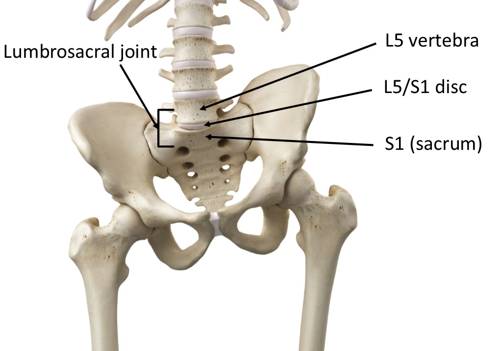
L5-S1 is the exact spot where the lumbar spine ends and the sacral spine begins. The lumbosacral joint is the joint that connects these bones. L5-S1 is composed of the last bone in the low back, called L5, and the triangle-shaped bone beneath, known as the sacrum. The sacrum is made of five fused bones, of which the S1 is the topmost.

Anatomical diagrams of the spine and back. This human anatomy module is composed of diagrams, illustrations and 3D views of the back, cervical, thoracic and lumbar spinal areas as well as the various vertebrae. It contains the osteology, arthrology and myology of the spine and back. It is particularly interesting for physiotherapists ...

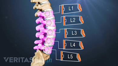
Mar 13, 2020 — Each lumbar spinal level is numbered from top to bottom—L1 through L5, or L6. The low back vertebral bodies are larger, thicker block-like ...

Christine Hudson Date: August 17, 2021 The extra vertebra is a defect in the lumbar vertebrae region of the spinal cord - a person will have a sixth vertebra as opposed to the normal five.. The term "extra vertebra" is most commonly used to describe an anatomical abnormality in which a person is born with six sacrum vertebrae in the spine instead of the more typical five.
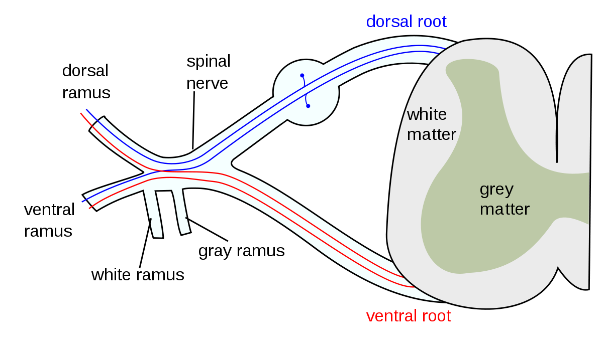
Dorsal branches, which give rise to the spinal branches that enter the vertebral canal. Supply. Dorsal muscles of the back, fascia, ligaments, vertebrae and intervertebral discs. Variant anatomy. A smaller fifth pair of lumbar arteries may be present, arising from the median sacral artery. The lumbar arteries may arise from a common trunk.

Radiograph of the lumbosacral junction showing a grade 1 spondylolytic spondylolisthesis at L5-S1. Lumbar oblique radiograph showing the "Scottie Dog." A pars defect is seen at L5.

(A) Schematic diagram showing the structure of the samples, data types, and flow of analyses in DIPPER. n is the number of individuals. N is the number of genome-wide profiles. (B) Clinical T2-weighted MRI images (3T) of the young lumbar discs in the sagittal and transverse plane (left and right panels), T1 MRI image of the young lumbar spine (middle panel).
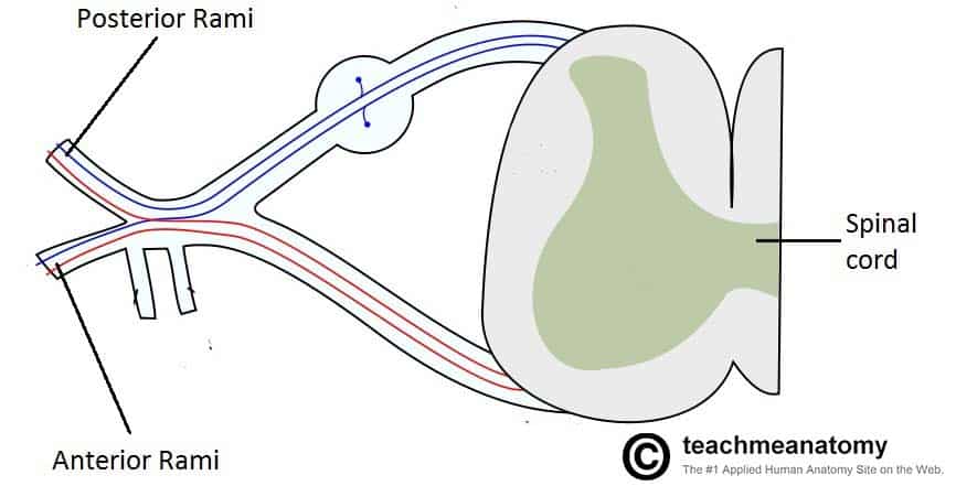
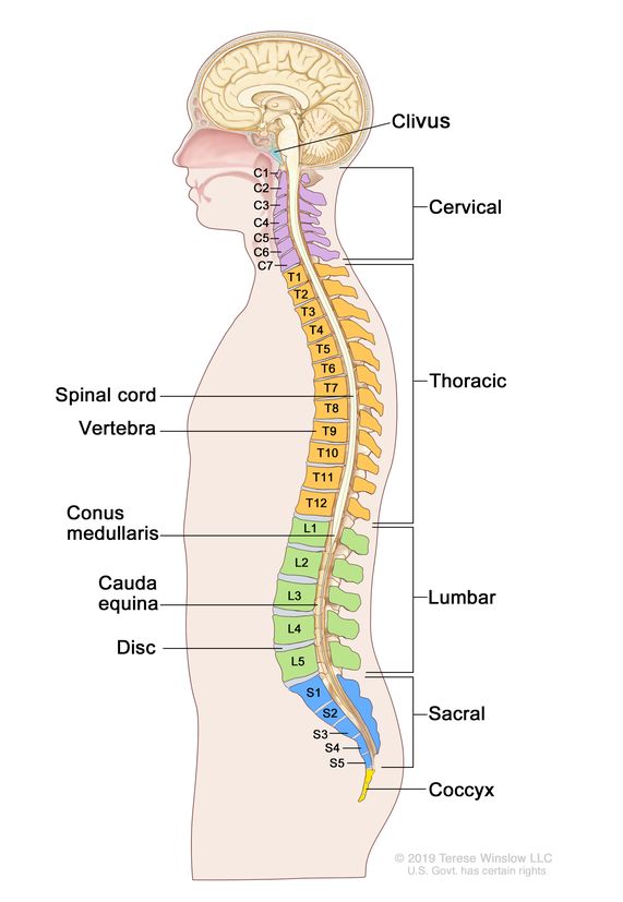
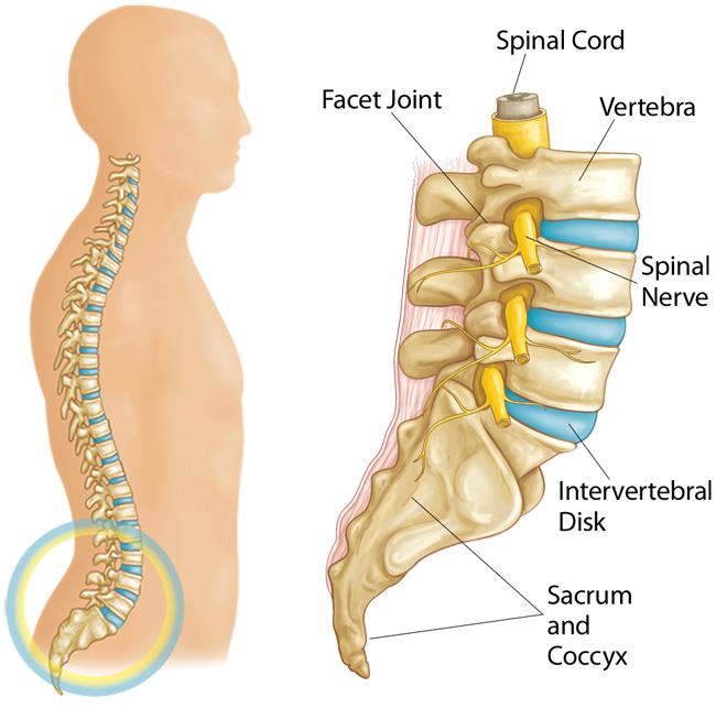
The spinal cord is protected by the spine, which is composed of 33 vertebrae. Peripheral nerves (which connect the central nervous system to the rest of the body) branch out from the spinal cord in pairs and exit above or below their corresponding vertebrae (C1-C7 spinal nerves exit above the corresponding vertebrae, while the rest of the spinal nerves from C8 downward all exit below).
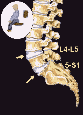
The L5-S1 spinal motion segment, also called the lumbosacral joint, is the transition region between the lumbar spine and sacral spine in the lower back.
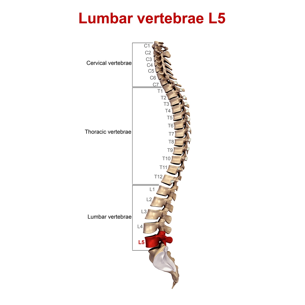
Five Regions of the Spine. As mentioned previously, the bony vertebrae of the spine are divided into cervical, thoracic, lumbar, sacral and coccygeal vertebrae. The sections are associated as ...
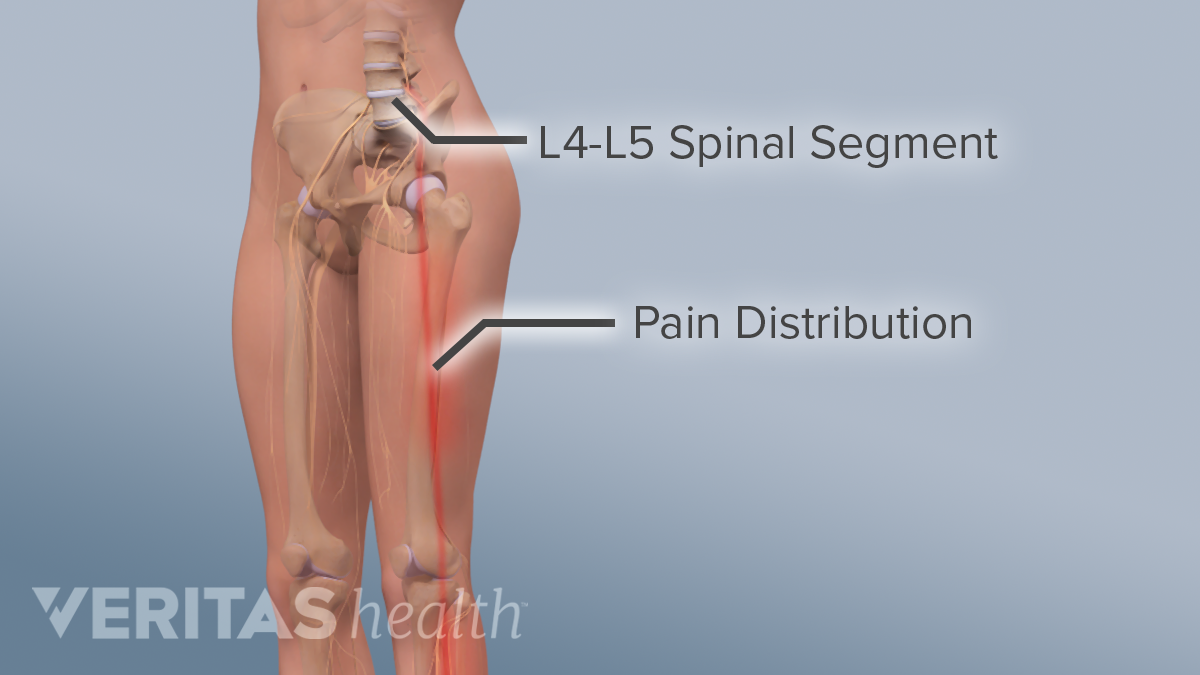
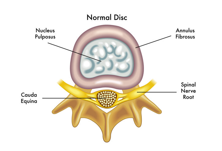
Lumbar Spinal Motion Segment · Two consecutive vertebrae, such as L4-L5, stacked vertically. · An intervertebral disc that has a soft inner gelatinous core ...
Lumbar (lower back), L1 to L5; Sacrum (buttocks), S1 to S5; Coccyx (buttocks) Our spine is made up of 33 individual bones known as vertebrae that are joined together to form the spinal column. The vertebrae are numbered individually and broken down into five sections:
the entire lumbar spine should be visible from T12/ L1- L5/S1 superimposition of the greater sciatic notches, the superior articulating facets and the superior and inferior endplates. This indicates a true lateral has been achieved

Surgical and nonsurgical management of lumbar spinal stenosis: four-year outcomes from the maine lumbar spine study. Spine (Phila Pa 1976) . 2000 Mar 1. 25(5):556-62. [Medline] .
Purpose The purpose of this study was to investigate and determine whether there are differences in L5 pedicles morphology between isthmic and degenerative L5-S1 spondylolisthesis. Methods One hundred and nineteen patients with isthmic spondylolisthesis and 45 patients with degenerative spondylolisthesis at L5-S1 were enrolled in the IS group and DS group, respectively, and 164 lumbar disc ...

740 lumbar spine anatomy diagram stock photos, vectors, and illustrations are available royalty-free. See lumbar spine anatomy diagram stock video clips. of 8. spinal vertebrae bone spine vertebra toracica spinal cord spine structure back diagram spine sections spinal cord vertebrae spinal structure health diagram. Try these curated collections.

The segment where the lumbar spine meets the sacral region, L5-S1, is an area that is prone to degenerate and create back problems. Four small bones that extend ...
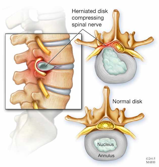
L5/S1 is known as a part of the spine that is under the most stress and also the most complex. It has distinctive anatomy which makes it a bit special. Sadly, this also means that this segment is most commonly affected by injuries which include degeneration and especially herniation. Herniated Disc L5/S1 may occur at any given moment to any person.
Anatomy of the lumbar spine (CT scan) This anatomy module is dedicated to interns and students that wish to learn more about the anatomy of the lumbar spine in CT. It allows to differentiate the vertebrae, the nervous system, the intervertebral discs and the zygapophyseal joints. The images are available in the three planes, axial, sagittal ...

Furthermore, we attached it along the spine, including S1-L1, L1/T12-C7, C1-C7, and S1-C7 (C, T, L, and S represent the cervical, thoracic, lumbar, and sacrum segments of the spine ...

Lastly is the sacrum, a large triangular bone at the base of the spine. It is formed by the fusing of sacral vertebrae S1-S5. Finding the Conus. You can either count from the sacrococcygeal area there will be 5 sacral and 5 lumbar vertebrae, the conus medullaris should end at of before L2.

/pelvis-joints-480791905-5bfd7b4b46e0fb0051b8ab26.jpg)


0 Response to "42 spine l5 s1 diagram"
Post a Comment