38 pacemaker cell action potential diagram
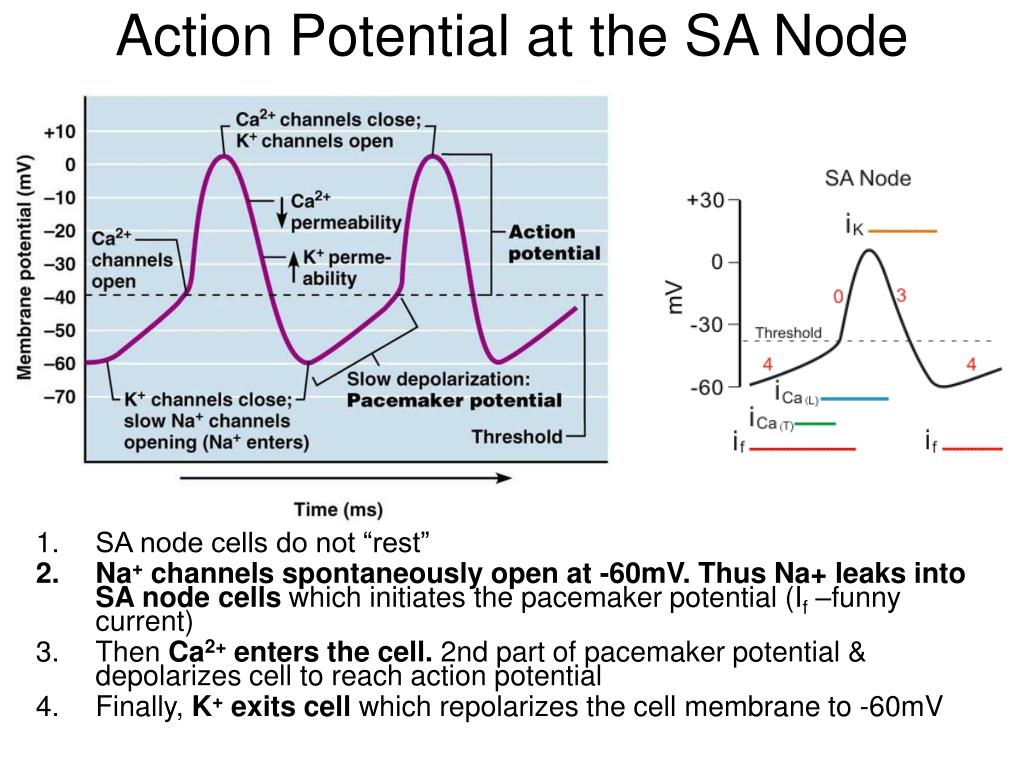
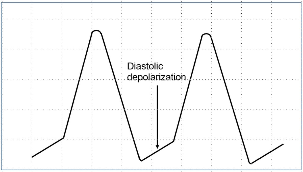
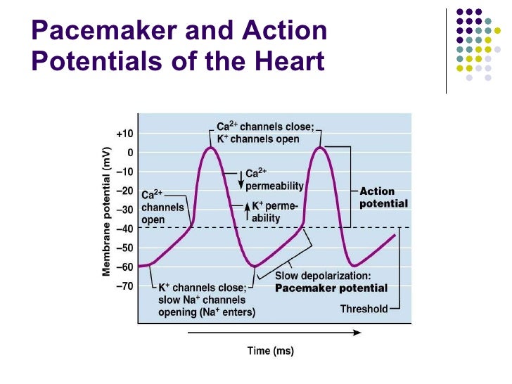
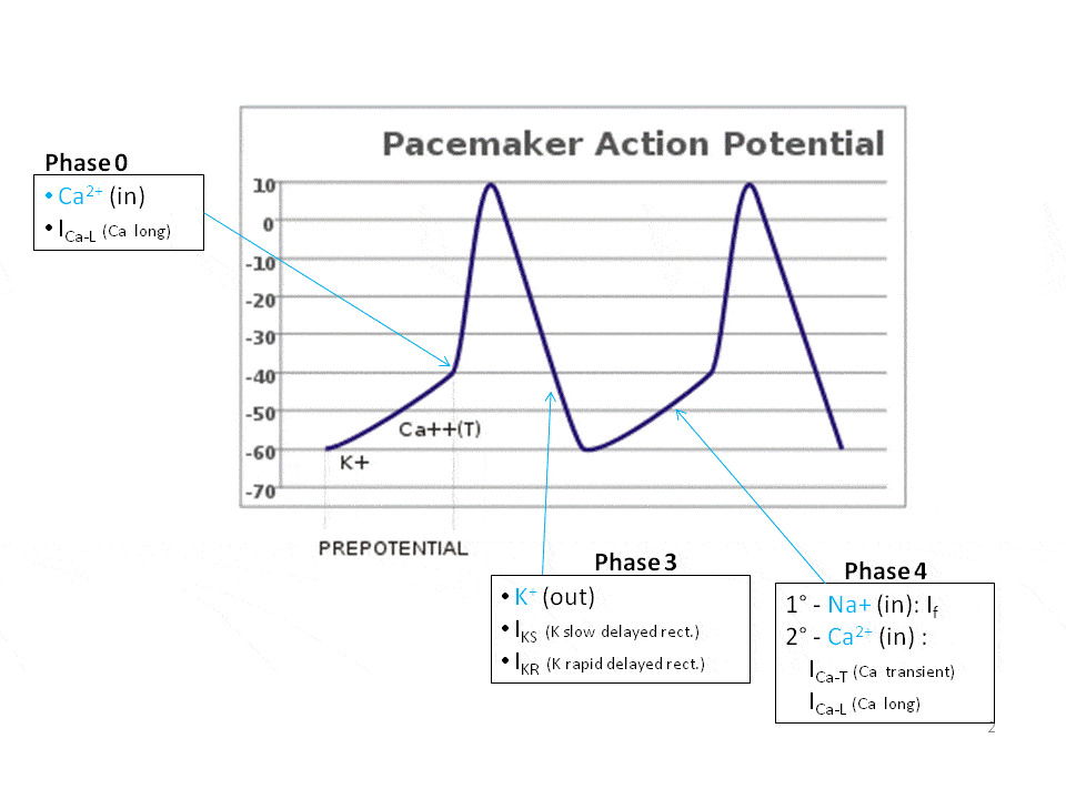
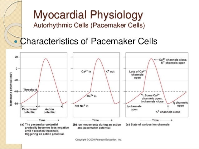
The action potential in the SA node occurs in three phases which are discussed below. The pacemaker potential occurs at the end of one action potential and just before the start of the next. It is the slow depolarisation of the pacemaker cells e.g. cells of the sinoatrial node, towards the membrane potential threshold. (a) Voltage changes in the heart as a function of changes in ion currents into and out of the cell. (b) Schematic of an ECG tracing. Parameters derived from the ...
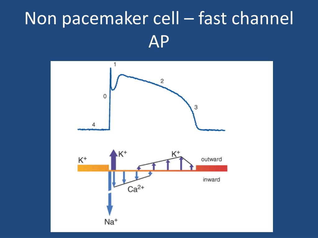
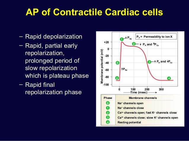
The above diagram is an action potential recorded in the ventricular cardiac cell of the heart. 4) Resting potentials in these cells is set by a large K+ permeability due to a combintation of the leak K+ channel and a voltage-gated K+ channel (called …

Pacemaker cell action potential diagram
Now, pacemaker cells also listen to which usually come from neighboring pacemaker cells. But if those don't come, then a pacemaker cell will simply launch its ...Jan 17, 2019 The action potential is then dispersed throughout the heart by myocardiocytes, cardiac muscle cells that contract while they conduct the current to neighboring cells. Similar to action potential initiation in neurons, and in contrast to pacemaker cells, myocardiocytes initiate rapid depolarization through voltage-gated sodium channels. Figure 17.2 Graph depicting the action potential of a pacemaker cell. ACTION POTENTIALS IN MYOCYTES osms.it/myocyte-action-potentials Myocytes Receive signal from from pacemaker cells causing them to contract Able to depolarize, spread action potentials Action potential phases: Phase 0 (depolarization phase): rapid influx of sodium into cell (inward current); responsible for rapid ...
Pacemaker cell action potential diagram. Download scientific diagram | The membrane action potential of pacemaker cells and the mechanism of action of several bradycardic drugs. In SA cells, when the membrane potential is repolarized to ... 1. Pacemaker Potential • An autorhythmic cell has the unique ability to depolarize spontaneously, resulting in a pacemaker potential. 2. Depolarization and Reversal of the Membrane Potential • Once threshold is reached, an action potential is initiated, which begins with further depolarization and leads to reversal of the membrane potential ... phase of depolarization occurs (Fig 2). At the peak of the action potential, K+ channels open, K+ rushes out of the cell and the cell repolarizes. Figure 2. Slow action potential has 3 phases (0, 3 and 4). The pacemaker cells set the rate of the heart beat. They are anatomically distinct from the The contraction of cardiac muscle (heart muscle) in all animals is initiated by electrical impulses known as action potentials.The rate at which these impulses fire controls the rate of cardiac contraction, that is, the heart rate.The cells that create these rhythmic impulses, setting the pace for blood pumping, are called pacemaker cells, and they directly control the heart rate.
This would support pacemaker cell excitability by preventing current dissipation during the slow diastolic and/or upstroke phase of pacemaker action potential generation. Thus, by creating a unique microenvironment, sinoatrial node fibrosis may provide direct biophysical support for pacemaker myocardium. Cells within the sinoatrial (SA) node are the primary pacemaker site within the heart. These cells are characterized as having no true resting potential, but instead generate regular, spontaneous action potentials.Unlike non-pacemaker action potentials in the heart, and most other cells that elicit action potentials (e.g., nerve cells, muscle cells), the depolarizing current is carried into ... Find out how the pacemaker cells use the movement of sodium, calcium, and potassium to get your heart beating! Rishi is a pediatric infectious disease physic... (b) Draw the action potential curves of a typical neuron, a pacemaker cell, and a cardiomyocyte, separately. Include all essential labeling, be careful with unit and legend. (c) How to identify whether a given action potential curve is obtained from a neuron, cardiomyocyte, or pacemaker cell, i.e. what are the characteristics of different cells ...
by S Kurbel · 2003 · Cited by 2 — They are also expected to draw a graph similar to Fig. 1. ... In SA cells, the resting membrane potential is never reached, because it lies ... ular node, and action potentials for non-pacemaker cells such as atrial or ventricular muscle cells. Pacemaker cells are capable of spontaneous action potential generation, whereas non-pacemaker cells have to be triggered by depolarizing currents from adja-cent cells. To compare non-oscillatory and oscillatory cells, it is convenient to rewrite Lusitropy is the rate of myocardial relaxation. The increase in cytosolic calcium of cardiomyocytes via increased uptake leads to increased myocardial contractility (positive inotropic effect), but the myocardial relaxation, or lusitropy, decreases. This should not be confused, however, with catecholamine-induced calcium uptake into the sarcoplasmic reticulum, which … For an easy explanation of pacemaker cell action potentials, make sure to check out the EZmed blog that makes cardiac action potentials easy! Once an action potential is generated by the SA node, it will travel though the right atrium via the internodal pathway, as well as to the left atrium via Bachmann's bundle.
The inherent pacemaker rate of the SA node is faster than the other pacemaker cells, and for that reason the SA node generates the initial action potential in a normal functioning heart. If the SA node becomes suppressed, then the other pacemaker cells are capable of generating spontaneous action potentials but at a slower heart rate.
length) A and B. In C recording is from a Intent or subsidiary pacemaker cell (upper) and true pacemaker (lower), rate of drive 300/min or 200 msec cycle length. Note the decrease in amplitude of pacemaker cell spike and the greater than normal duration of pacemaker cell action potentials after drive.
A paraganglion (pl. paraganglia) is a group of non-neuronal cells derived of the neural crest.They are named for being generally in close proximity to sympathetic ganglia.They are essentially of two types: (1) chromaffin or sympathetic paraganglia made of chromaffin cells and (2) nonchromaffin or parasympathetic ganglia made of glomus cells.They are neuroendocrine …
(USMLE topics, cardiology) Cardiac action potential in pacemaker cells and contractile myocytes, electrophysiology of a heartbeat. This video is available fo...
Nodal Cell Action Potential Profile. This diagram illustrates the action potential profile of a nodal cell within the heart, with time plotted against membrane potential. From this diagram, several characteristics of a nodal cell can be noted. ... Pacemaker cells do not compete, they go at the fastest rate. In a healthy human, this is the SA node.
The action potential for the conductive cells consists of a prepotential phase with a slow influx of Na + followed by a rapid influx of Ca 2+ and outflux of K +. Contractile cells have an action potential with an extended plateau phase that results in an extended refractory period to allow complete contraction for the heart to pump blood ...
Cardiac Myocyte Action Potential. This diagram is a diagram of a cardiac myocyte - a ventricular muscle cell as apposed to a cardiac pacemaker cell. The resting membrane potential (RMP) is -90mv. A membrane potential is the difference in electrical potential between the interior and the exterior of the cell membrane.
Pacemaker potential. In the pacemaking cells of the heart (e.g., the sinoatrial node ), the pacemaker potential (also called the pacemaker current) is the slow, positive increase in voltage across the cell 's membrane (the membrane potential) that occurs between the end of one action potential and the beginning of the next action potential.
Figure 1. The action potential in the sinoatrial node and in contractile myocardial cells. Phase 4 of the action potential in the sinoatrial node is called 'pacemaker potential', because it is responsible for the spontaneous repetitive depolarization. The depolarization spreads from the sinoatrial node to the atrial and ventricular myocardium.
Figure 17.2 Graph depicting the action potential of a pacemaker cell. ACTION POTENTIALS IN MYOCYTES osms.it/myocyte-action-potentials Myocytes Receive signal from from pacemaker cells causing them to contract Able to depolarize, spread action potentials Action potential phases: Phase 0 (depolarization phase): rapid influx of sodium into cell (inward current); responsible for rapid ...
The action potential is then dispersed throughout the heart by myocardiocytes, cardiac muscle cells that contract while they conduct the current to neighboring cells. Similar to action potential initiation in neurons, and in contrast to pacemaker cells, myocardiocytes initiate rapid depolarization through voltage-gated sodium channels.
Now, pacemaker cells also listen to which usually come from neighboring pacemaker cells. But if those don't come, then a pacemaker cell will simply launch its ...Jan 17, 2019












0 Response to "38 pacemaker cell action potential diagram"
Post a Comment