39 diagram of synovial joints
The interphalangeal joints of the hand also known as Bitals, are the hinge joints between the phalanges of the fingers that provide flexion towards the palm of the hand.. There are two sets in each finger (except in the thumb, which has only one joint): A synovial joint is characterised by the presence of a fluid-filled joint cavity contained within a fibrous capsule. It is the most common type of joint found in the human body, and contains several structures which are not seen in fibrous or cartilaginous joints.. In this article we shall look at the anatomy of a synovial joint - the joint capsule, neurovascular structures and clinical ...
Synovial Joint Diagram Labeled Anatomy Chart With Two Bones Synovial joints are characterized by the presence of a joint cavity. It consists of two layers. Fine in this article I will describe the synovial joint structure with a labeled diagram. Allows movement in only one plane. Tough Fibrous tissue surrounds synovial joints.
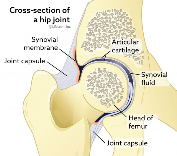
Diagram of synovial joints
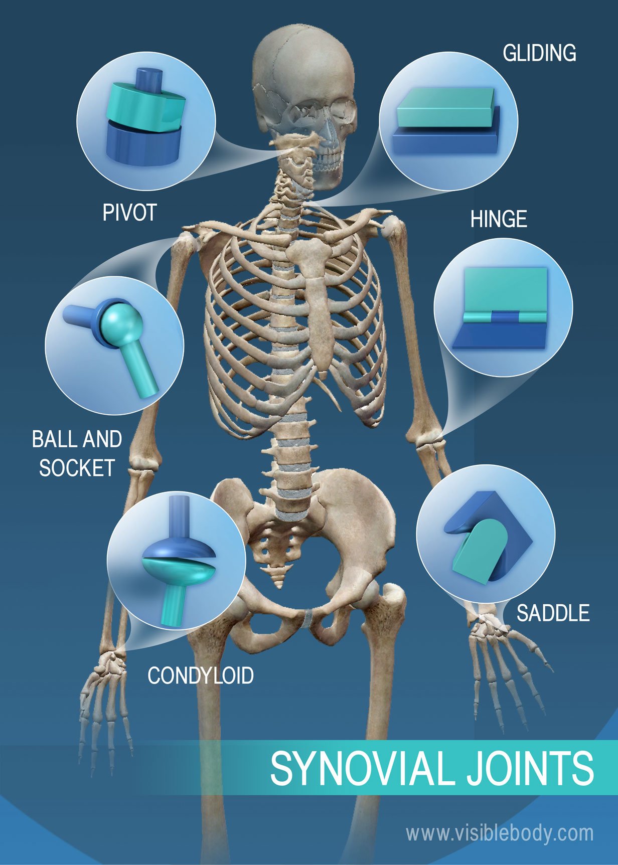
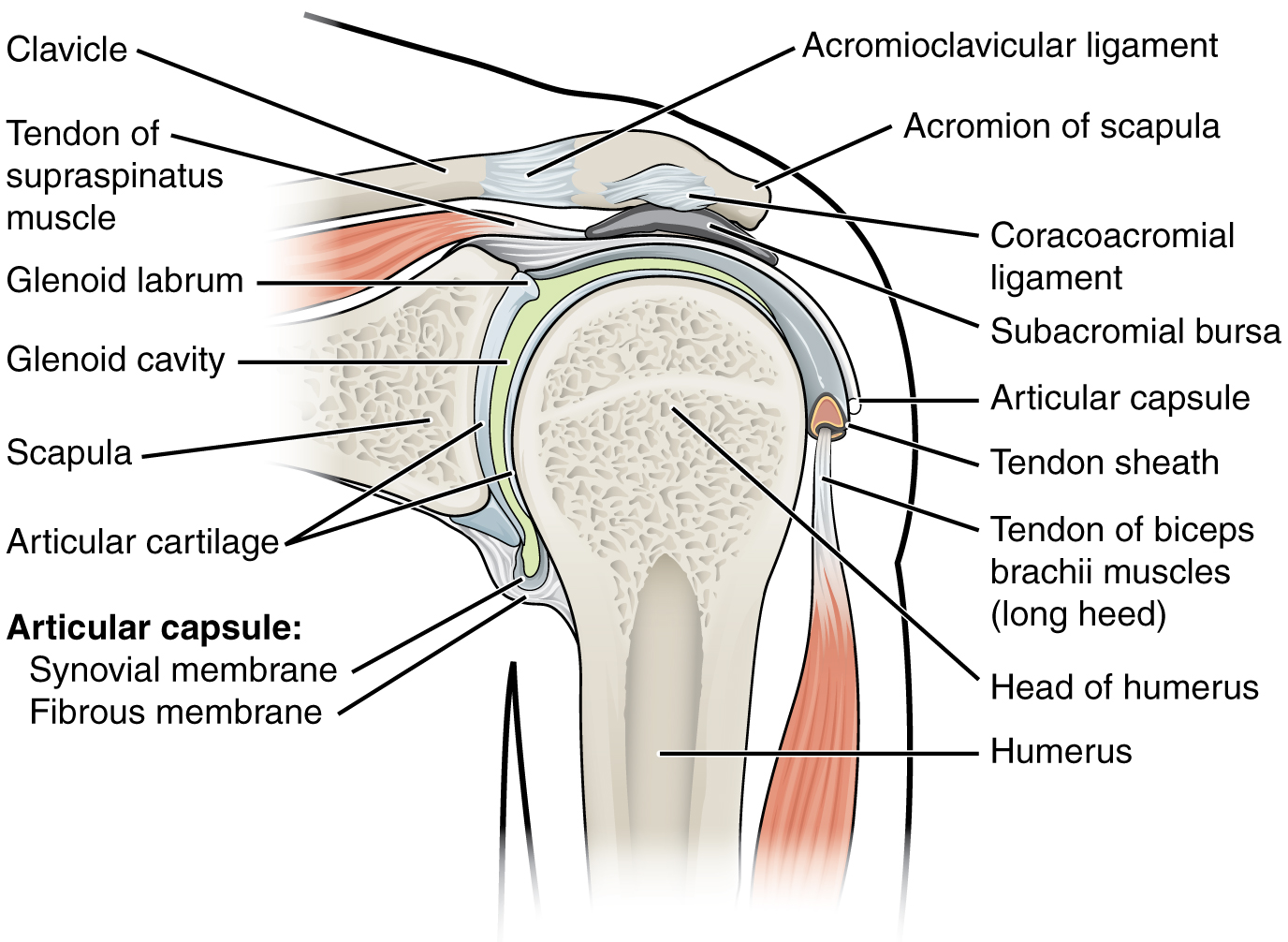
Sep 16, 2021 · The biceps works across three joints. The most important of these functions is to supinate the forearm and flex the elbow. Besides, the long head of the biceps prevents the upward displacement of the head of the humerus. 3. Label the six different types of synovial joints on the diagram by clicking and dragging the labels to the correct location Saddle Joint bok Hinge joint ences 4 Pivot joint Ball and socket N 5 Condylar joint 3 6 Plane (gliding) joint ; Question: 3. Label the six different types of synovial joints on the diagram by clicking and dragging the ... Classification of Joints • 1. According to the type of tissue at the joint: • a) Fibrous joint -- uses fibrous connective tissue to articulate bones. • b) Cartilaginous joint-- uses hyaline cartilage and/or fibro- cartilage to articulate bones. • c) Synovial joint --uses auricular cartilage, synovial membrane, joint capsule, and ligaments to articulate bones.
Diagram of synovial joints. Synovial joint diagram Here in the diagram, you will find all the structures of a synovial joints in animals. If you need more diagram like this synovial joint then, you may follow anatomy learner blog or social media. Again, you might read other different article related to veterinary osteology or syndesmology with the anatomy learner. Synovial joints may also become inflamed, called arthritis. There are more than 100 different types of arthritis, arising from problems in different parts of the joint. For example in osteoarthritis, the cartilage becomes worn , and in rheumatoid arthritis the body's immune system attacks the synovial membrane. CARTILAGINOUS JOINTS Hyaline cartilage connects bones, stretches to allow some movement Synchondrosis: costochondral joint, where cartilage attaches rib to sternum; growth plates between bone diaphysis, epiphysis Symphysis: symphysis pubis in pelvic bone (fibrous cartilage) ↑ strength, ↓ flexibility SYNOVIAL JOINTS Joint capsule connects ... The temporomandibular joint (TMJ), or jaw joint, is a synovial joint that allows the complex movements necessary for life. It is the joint between condylar head of the mandible and the mandibular fossa of the temporal bone. This system is made up of the TMJ, teeth and soft tissue and it plays a role in breathing, eating and speech.
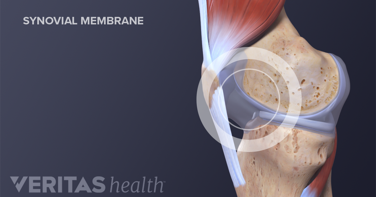
Nov 3, 2013 - synovial joint. Easy pic for patients to understand and you talk about joint health. Nov 3, 2013 - synovial joint. Easy pic for patients to understand and you talk about joint health ... Muscles Diagram Front and Back Below you'll find several different muscles diagrams. There are anterior muscles diagrams and posterior muscles ... Next, let's focus on hinge joints, shown as letter B on the diagram. Hinge joints are the synovial joint type referred to in our introductory section. These joints can be found between your upper ... The only 2 synovial joints that aren't diarthrotic. carpals and tarsals. Functional classification of carpals and tarsals. amphiarthrosis. membrane continuous from bone to bone outside the articular capsule. periosteum. fibrous capsule lined by synovial membrane. articular capsule. Labelled Diagram Of Synovial Joint. A synovial joint is a connection between two bones consisting of a cartilage lined As seen in the above picture, the most powerful bite in the world gets its. A synovial joint or diarthrosis occurs at articulating bones to allow movement. fibrous connective tissue found in various parts of the body such as ...
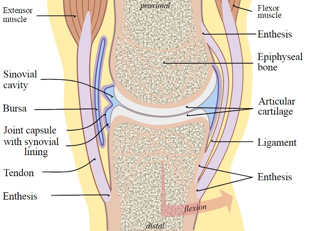
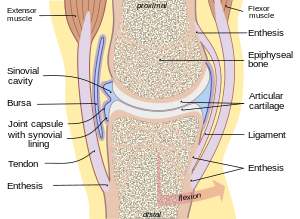
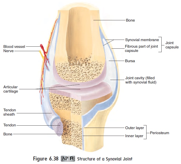
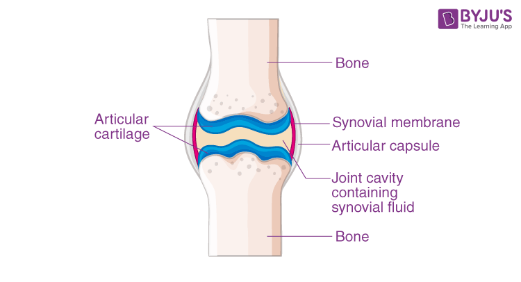
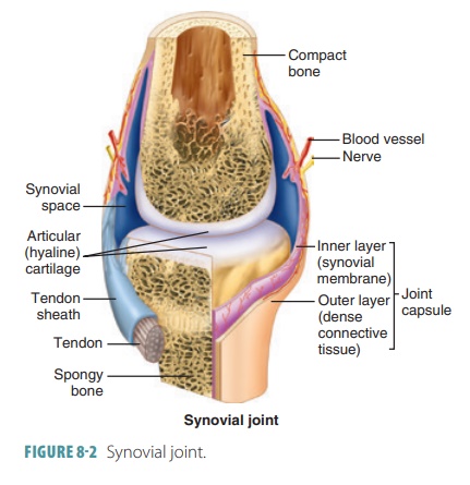
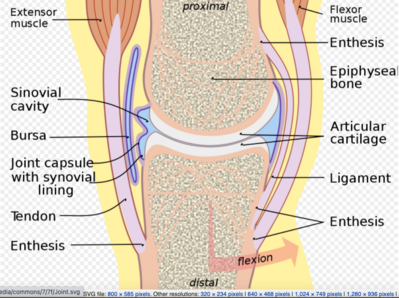
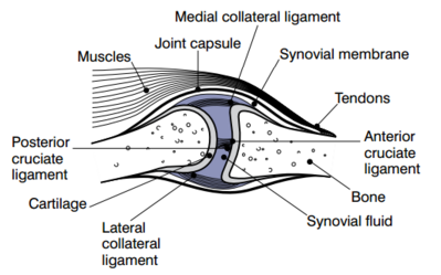
The synovial cavity/joint is filled with synovial fluid. The joint capsule is made up of an outer layer, the articular capsule, which keeps the bones together ...Latin: junctura synovialisTA2: 1533 Components of synovial jointsComponents of synovial joints •A jjpoint capsule consists of two layers - Fibrous capsule holds the ends of the bones together and allows movement of the joint - Synovial membraneSynovial membrane which consists of connectivewhich consists of connective tissue • Synovial fluid: combination of materials filtered from blood and secreted by cells of the synovial The synovial joint is a moveable or true joint in an animal's body. Hi there, do you want to learn synovial joint anatomy in animals? Fine, in this article, I will describe the synovial joint structure with a labeled diagram. I will also describe different types of synovial joints in animals. After reading this article, you will know the ... Synovial Joints. Joints can be simply defined as articulations of bones, which functions by providing shape to the skeleton system, protects bones by holding them together securely and also helps in movement. Based on structure and functions, joints have been further classified into different types. A synovial joint is one among the three types ...
Degenerative Joint Disease In Dogs | VCA Animal Hospitals
Note: Synovial joint joins bones or cartilage with a fibrous joint capsule which continues with the periosteum of the bone, and forms the outer boundary of a ...1 answer · Top answer: Hint: A synovial joint is the type of joint found between two bones that move against each other, such as the joints of the limbs (e.g. shoulder, hip, elbow ...
Solved] Use the list of structures provided to label the ...
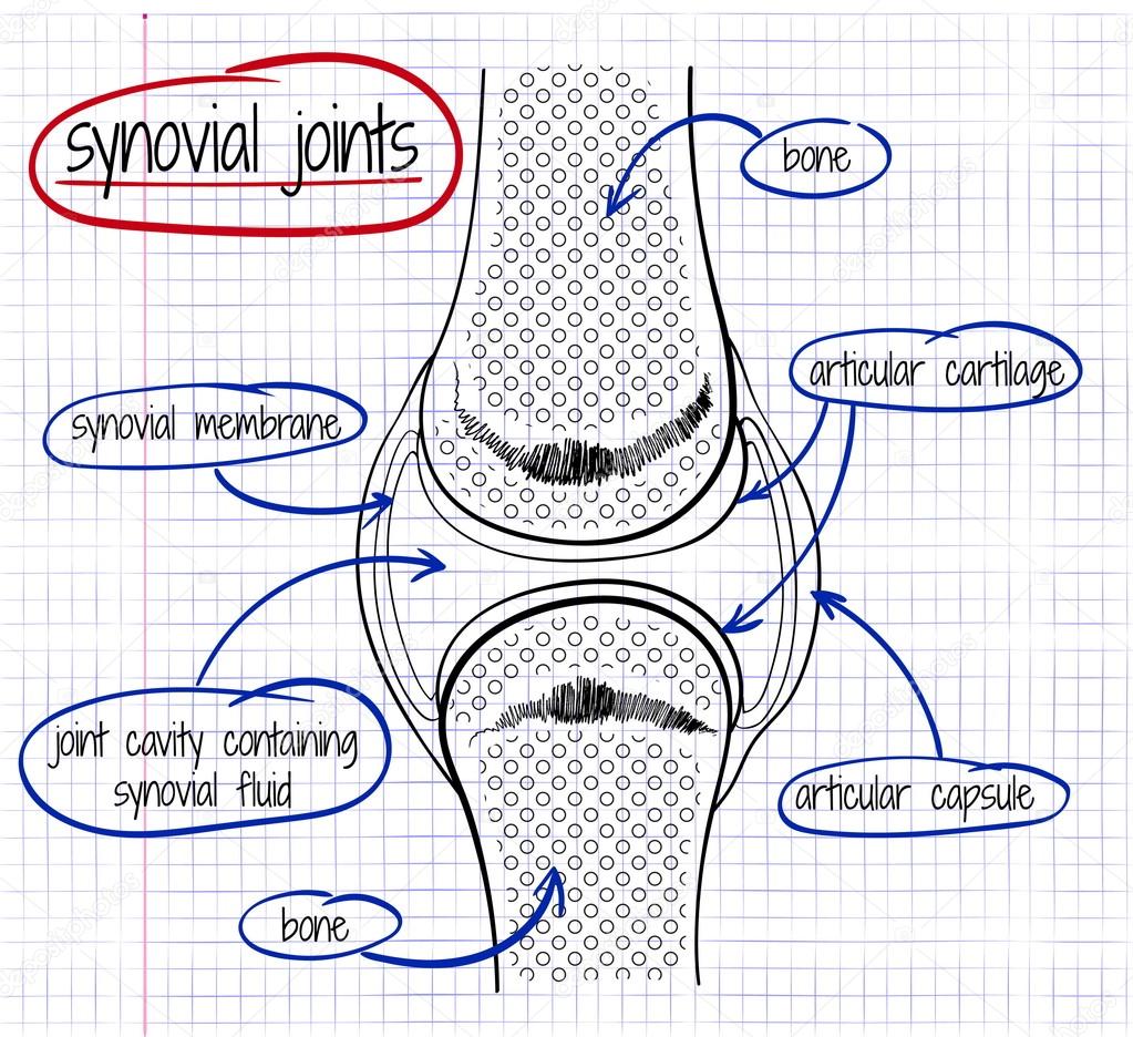
The main parts of synovial joints are labelled on the synovial joint diagram and described in the table below. A, The egg-shaped ovoid surface represents a characteristic of most synovial joints of the body (e.g., hip joint, radiocarpal joint, knee joint, metacarpophalangeal joint). The diagram shows only the convex member of the joint.
Synovial joint | Radiology Case | Radiopaedia.org
Four important synovial joints The elbow and knee joints are both hinge joints. A hinge joint is a type of synovial joint that works like the hinge on a door, allowing bending and straightening only.

Synovial Joint Diagram | Quizlet
Oct 07, 2021 · Costovertebral joints and ligaments: Diagram The two joints are reinforced by three costotransverse ligaments (medial, lateral, superior). These run from the transverse processes to the neck and tubercle of the ribs, respectively. In addition, intra-articular and radiate ligaments of head of rib also support these joints. These extend to the ...
What Is a Synovial Joint?
A synovial joint is also called diarthrosis, joint cartilage or bones with a fibrous joint. These joints allow bones to rotate around each other and slide past each other. The synovial joint has a joint cavity filled with fluid, together with muscles, ligament, tendons, and the capsule which keeps the bones of the joint in place.

How To Draw The Structure Of synovial Joint. VERY SIMPLE
A worksheet that covers a diagram of each synovial joint and a table to complete the description and locations of this joint type in the body Nice easy task for a lesson on the skeletal system. Subjects: Anatomy, Biology, Health. Grades: 6 th - 12 th. Types: Activities, Handouts, Worksheets.
Joints and Ligaments | Learn Skeleton Anatomy
What type of synovial joint is shown in the diagram? Condyloid. Plane. Hinge. Saddle. Ball and Socket. Pivot. The Elbow Joint: Functions And Location! Quiz . The Elbow Joint: Functions And Location! Quiz. How much do you know about the elbow joint, functions, and location? The elbow is a visible joint between the upper and lower parts of the arm.
Joint - Wikipedia
Synovial Joint. Create healthcare diagrams like this example called Synovial Joint in minutes with SmartDraw. SmartDraw includes 1000s of professional healthcare and anatomy chart templates that you can modify and make your own. 26/37 EXAMPLES.
Joints
Start studying types of synovial joints (ex. 11). Learn vocabulary, terms, and more with flashcards, games, and other study tools.

a) A Synovial Joint; (b) Types of Synovial Joints. Image ...
Next, let's focus on hinge joints, shown as letter B on the diagram. Hinge joints are the synovial joint type referred to in our introductory section. These joints can be found between your upper and lower arm bones, otherwise called your elbow, as well as your ankles, fingers, toes, and knees. Hinge joints operate just like the hinges on a door.

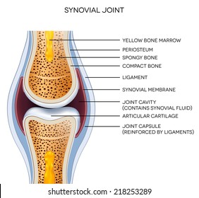
Figure 8.3 General structure of a synovial joint.
Describe the structure of synovial joint with the help of a neat labelled diagram. Hard. Open in App. Solution. Verified by Toppr. When the two bones are joined with the help of connective tissues which allows the movement of the bones is known as the synovial joint. This is a joint which is present between the long bones. The two bones are ...
Draw A Labelled Diagram Of A Synovial Joint. Give Examples ...
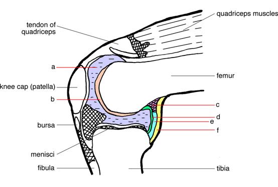
Synovial joints allow for smooth movements between the adjacent bones. This diagram shows the location of the bursae which are fluid filled sacs in a bone. The basic structure of a synovial joint is shown in the diagram below. The main parts of synovial joints are labelled on the synovial joint diagram.

Joints | BioNinja
The structure of a synovial joint is demonstrated by a diagram in which the articulating bones are surrounded by the articular capsule, which comprises an exterior fibrous capsule and an interior synovial membrane. Start studying label the synovial joint. Learn vocabulary, terms, and more with flashcards, games, and other study tools.

Describe typical synovial joint with a neat labelled diagram ...
Synovial joints are subdivided based on the shapes of the articulating surfaces of the bones that form each joint. The six types of synovial joints are pivot, hinge, condyloid, saddle, plane, and ball-and socket-joints ( Figure 9.4.3 ).

synovial joint. Easy pic for patients to understand and you ...
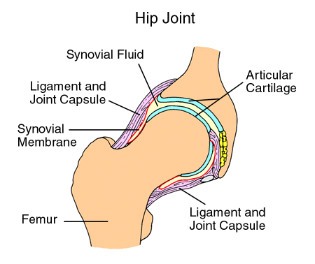
The synovial fluid is similar to raw egg whites in its consistency. The function of the bursae and the synovial fluid is to reduce the friction that occurs as ligament and bones, ligaments and tendons, or tendons and bones rub together. Joints have bursae around them, and because the hip joint is so large it has many bursae, around 20 total.

Structure of synovial joint
Synovial joints. A joint is a place where two or more bones meet and is also called an articulation. The role of joints and connective tissue . Connective tissues consist of ligaments, cartilage ...

Types Of Synovial Joints! Trivia Questions Quiz - ProProfs Quiz
Classification of Joints • 1. According to the type of tissue at the joint: • a) Fibrous joint -- uses fibrous connective tissue to articulate bones. • b) Cartilaginous joint-- uses hyaline cartilage and/or fibro- cartilage to articulate bones. • c) Synovial joint --uses auricular cartilage, synovial membrane, joint capsule, and ligaments to articulate bones.

Osteoarthritis Healthy Synovial Joint And Knee With Arthritis ...
3. Label the six different types of synovial joints on the diagram by clicking and dragging the labels to the correct location Saddle Joint bok Hinge joint ences 4 Pivot joint Ball and socket N 5 Condylar joint 3 6 Plane (gliding) joint ; Question: 3. Label the six different types of synovial joints on the diagram by clicking and dragging the ...

Synovial Joint worksheet
Sep 16, 2021 · The biceps works across three joints. The most important of these functions is to supinate the forearm and flex the elbow. Besides, the long head of the biceps prevents the upward displacement of the head of the humerus.
2.2.4 Anatomy of Selected Synovial Joints – Biomechanics of ...

Synovial Joints: Structure, Function & Types | Study.com
Synovial Joints
Anatomy of a Joint | Children's Wisconsin

Draw labelled diagram Synovial joint. - Biology | Shaalaa.com

Articulations
Synovial joint activity
Synovial joint Images, Stock Photos & Vectors | Shutterstock

How to Prevent Osteoarthritis - Cathedral Chiropractic

Types of Synovial Joints | Biology for Majors II

9.2: Diarthroses: Synovial joints contain synovial fluid and ...

تويتر \ Zeph Bennett على تويتر: "Labelled synovial joint ...
Synovial Joints - Physiopedia

Joints | BioNinja
Synovial Joints - Physiopedia

Synovial fluid in joints: what it is and how exercise affects ...
Vector drawing of a synovial joint Stock Vector Image by ...
Synovial Joint Structure - TeachPE.com

Synovial Joint - an overview | ScienceDirect Topics

Synovial (movable) Joints
0 Response to "39 diagram of synovial joints"
Post a Comment