41 arteries of the brain diagram
22 Oct 2021 — The arterial blood supply to the brain can be divided into the anterior and posterior circulation. The former is derived from the left and right ... 6 Aug 2013 — Intracranial arteries are involved in many neurologic disorders. Knowledge of arterial anatomy, variants, and areas involved in disease is ...
The mid brain has some key structures of the brain. Pontine Arteries: These small arteries branch off the Basilar Artery and supply the pons and other local parts of the brain with brain. Superior Cerebellar Artery: This vessel separates itself from the Posterior Cerebral Artery and passes by the cerebellum to supply the pia matter and areas around it with blood. Anterior Spinal Artery: The ...

Arteries of the brain diagram
Peripheral artery bypass - leg. Peripheral artery bypass is surgery to reroute the blood supply around a blocked artery in one of your legs. Fatty deposits can build up inside the arteries and block them. A graft is used to replace or bypass the blocked part of the artery. The graft may be a plastic tube, or it may be a blood vessel (vein ... Cached. Jan 03, 2022 · The developing blood vessel to the heart muscle does not attach correctly. In the normal heart, the LCA originates from the aorta. It supplies oxygen-rich blood to the heart muscle on the left side of the heart as well as the mitral valve (the heart valve between the upper and lower chambers of the heart on the left side). It keeps the diastolic (minimum) and systolic (maximum) pressure in the arteries under normal limits. In the case the blood pressure rises beyond the bearable limits, you are very likely to suffer from a heartattack, brain haemorrhage or other critical circulatory disorders.
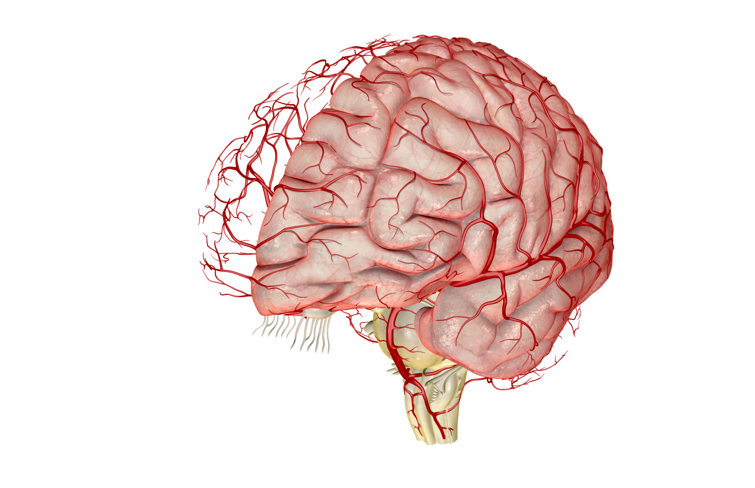
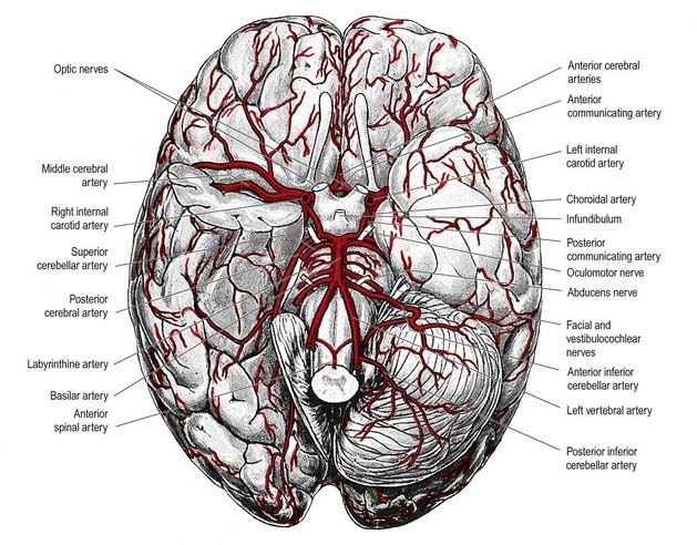
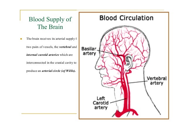
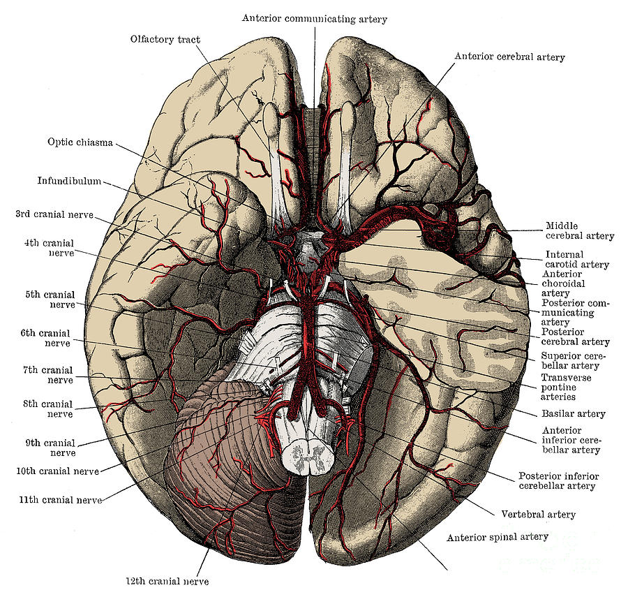
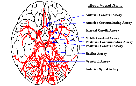
Arteries of the brain diagram. Anatomy Of The Cerebral Vasculature Diagram In this image, you will find cerebral arteries, cerebral artery, right anterior cerebral artery, anterior communicating artery, middle cerebral artery, internal carotid artery, posterior communicating artery, left anterior cerebral artery, left anterior cerebral artery A1 in it. Thunder claps are shock waves created by the rapid expansion of air following the formation of lightning. As it propagates, the shock waves attenuate, reflect and overlap, resulting in the transformation of thunder from a sharp clap to a low rumble. Lightning and the rumbling sound of thunder have intrigued and enticed humans for centuries. A total of 10 arteries were evaluated including OA, ACA, MCA, anterior choroidal artery (AchA), posterior choroidal artery (PchA), peri-callous artery (PcaA) and posterior cerebral artery (PCA), superficial temporal artery (STA), middle meningeal artery (MMA) and occipital artery (OcciA). The internal carotid arteries supply oxygenated blood to the front of the brain and the vertebral arteries supply blood to the back of the brain. These two circulations join in the circle of Willis, a ring of connected arteries that lies in the interpeduncular cistern between the midbrain and pons.
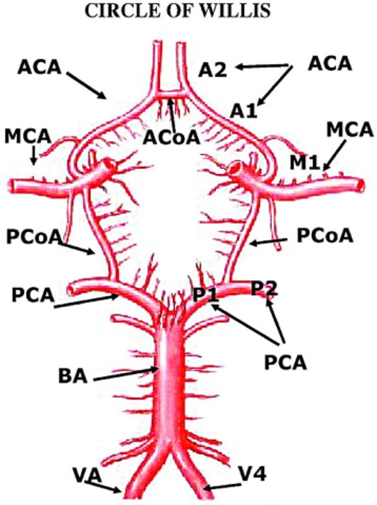
It is formed in front by the anterior cerebral arteries, branches of the internal carotid, which are connected together by the anterior communicating; behind by the two posterior cerebral arteries, branches of the basilar, which are connected on either side with the internal carotid by the posterior communicating (Figs. 516, 519). The auxiliary arteries, which delivers blood to your upper extremities The descending aorta, which delivers blood to the lower parts of the body. As blood is delivered to the organs and tissues, it passes through smaller blood vessels called capillaries, where it can easily distribute nutrients and oxygen, while removing wastes and carbon ... Dec 31, 2021 · Pulmonary arteries take blood with low levels of oxygen from the right ventricle to the lungs. ... The brain is divided into the right and left hemisphere, and the two halves are connected by the ... Brain Pathol. 2015 Jan ... Congo red staining of a small cortical artery at 400× magnification demonstrates salmon-colored amyloid deposition in the media of the vessel. ... This Venn diagram ...
January 4, 2022 Posted by Samanthi. The key difference between lymphangitis and lymphadenitis is that lymphangitis is the inflammation of lymphatic channels due to the infection at a site distal to the channel, while lymphadenitis is the enlargement of one or two lymph nodes due to an infection. The lymphatic system is a network of tissue ... A narrowed part between splenium and trunk is known as the isthmus. The corpus callosum always needs a constant and abundant blood supply to perform its functions. Infarcts uncommonly involve it. Mostly it gets its blood supply via pericallosal, the posterior pericallosal arteries, and branches from the anterior and posterior cerebral. Fig 1.3 – Schematic of the blood supply to the brain. Circle of Willis Regional Blood Supply to the Cerebrum There are three cerebral arteries; anterior, middle and posterior. They each supply a different portion of the cerebrum. The anterior cerebral arteries supply the anteromedial portion of the cerebrum. Throat Anatomy. Last reviewed by Dr. Raj MD on August 13th, 2018. The throat is a complex part of the body with many structures that surround it that are very important. In this article, you will read about each specific area of the throat as well as its surrounding structures. The throat is positioned in the anterior part of the neck.
Circulation and the Central Nervous System | Anatomy and ...
There are two types of them: arteries and veins. Arteries: These types of blood vessels take oxygen-rich blood from the heart and transport to the capillaries. Arteries are quite tough on the outside but are smooth on the inside. There are three arteries of the heart, including pulmonary artery, aorta, and coronary arteries.

Image from page 304 of "The structure and life of birds" (1895)
The connection between the brain and the heart was first noticed in the literature in 1846 by Sir George Burrows, who described the relationship between heart diseases and disorders of the cerebral circulation [].A few years later, Carl Ludwig announced the discovery of the depressor and accelerator nerves of the heart.

Image from page 544 of "Anatomy, descriptive and surgical" (1887)
The brain stem receives its blood supply exclusively from the posterior circulation, including the vertebrae and basilar artery. The medulla receives its blood supply from the vertebral via medial and lateral perforating arteries. The pons and midbrain receive their blood from the basilar via the medial and lateral perforating arteries.

water and the human body
The Demon core is a 6.2 kg Plutonium-Gallium sphere with a diameter of 8.9 cm (smaller than a basketball). The Plutonium-239 isotope used in the core is a manmade element produced in nuclear reactors. It is highly unstable, radioactive, and decays by emitting alpha particles, which is damaging to our tissues. Hence, the core has a nickel coating, which blocks the short-range alpha particles ...

Common variants of the circle of Willis: diagrams | Image ...
The artery is separated into two parts known as the right and left gastroepiploic arteries. Its point of origin is usua. Arteries Diagram / ANAT2241 Liver, Gallbladder, and Pancreas - Embryology - The bulbourethral artery is a short, relatively wide blood vessel found only in males that.. These parts of the body include the thorax, upper limb ...
Closeup of skeleton pelvic model
Looks like a sea-horse . The occipital lobe is located at the back portion of the brain and is associated with vision. We can learn a lot about the relationship between the brain and circumstances allowed to complete, deface or alter the diagram or narrative in any way. 5.

Amazing architecture of the Lou Ruvo Centre in Las Vegas, Nevada.
Systemic lupus erythematosus (SLE) is a systemic autoimmune disease characterized by multiple immunologic abnormalities and has the potential to involve the central nervous system (CNS). The prevalence of SLE seems to be growing, possibly because of earlier diagnosis and improved survival; however, the associated mortality is still high. The mortality is associated with disease-related risk ...
Migraines Linked With Abnormal Blood Vessel Structure In ...
The brain receives blood from two sources: the internal carotid arteries, which arise at the point in the neck where the common carotid arteries bifurcate, and the vertebral arteries (Figure 1.20). The internal carotid arteries branch to form two major cerebral arteries, the anterior and middle cerebral arteries .

Circle of Willis - Location, Anatomy, Function and FAQs
Insectivorous plants trap insects and other smaller animals to derive nutrition for themselves. The mechanism of capturing their prey differs among species, with some using rapid movements and others using slow movements. Insectivorous plants, also known as carnivorous plants, have been a marvel for both scientists and common people for eons.

Pin on Ανατομία
The brain is supplied by branches of the internal carotid artery anteriorly and by branches of the vertebral artery posteriorly. The aortic arch gives off three great vessels: the brachiocephalic artery, the left common carotid artery and the left subclavian artery.

Brainstem vasculature and emerging cranial nerves (A ...
Cerebral small vessel disease (cSVD), which encompasses a group of pathological processes that affect small penetrating vessels (arterioles, capillaries and venules) of the brain 2, 3, makes a ...

Pin on Anatomy/PT
A blood clot in the brain can cause an ischemic stroke which is caused by an artery to the brain becoming blocked and starving a portion of the brain of oxygen and nutrients. A clot in the brain can also cause a buildup of cellular waste and carbon dioxide because the brain will be unable to clear this waste properly.

Image from page 303 of "Some points in the surgery of the brain and its membranes" (1907)
Jul 23, 2021 · The Brain Hemispheres: Left Brain/Right Brain Communication and Control 7:06 Parts of the Brain Stem: The Medulla Oblongata and Pons 6:27 Brain Blood Supply: Anatomy & Diagrams 10:37
Print Exercise 32: Anatomy of Blood Vessels flashcards ...
Spinal fluid containing spaces in the brain 3- Bleeding inside the coverings of the brain Subarachnoid hemorrhage SAH - 85 related to aneurysms balooning of brain arteries. Intraparenchymal hemorrhage also called intracerebral hemorrhage refers to nontraumatic bleeding into the brain parenchyma Figure 2. What are the symptoms of brain hemorrhages.
Brain Aneurysm Symptoms & Treatment | Pacific Stroke ...
Fishbone diagrams are the versatile cause-and-effect diagrams that can help uncover the root causes of issues from the very granular. The Fishbone Diagram Examples. The first company to adopt the diagram in all of its processes was. The fishbone diagram also known as the Ishikawa diagram is a cause and effect diagram.
![7 Arteries of the brain [110]. | Download Scientific Diagram](https://www.researchgate.net/profile/Samara_Alzaidi/publication/38319379/figure/fig13/AS:669513129406471@1536635777310/The-ACA-arteries-31_Q320.jpg)
7 Arteries of the brain [110]. | Download Scientific Diagram
All of these responses to change the body start in the brain and gets sent out via chemicals or electrical messengers. The negative feedback loop helps to balance homeostasis by recognizing there is a problem in the body and sending out the right response. The loop stays where it is at and sends out different "effectors" to do the work.
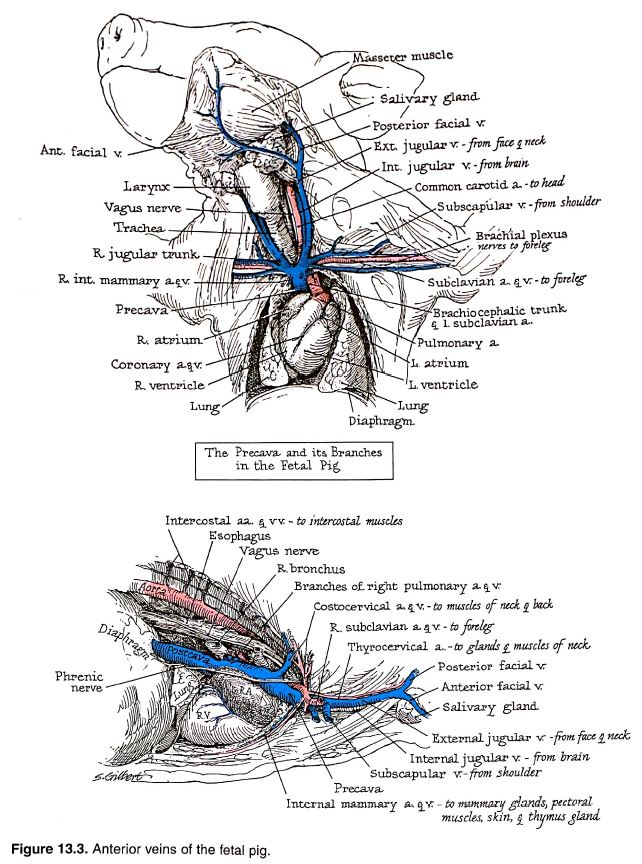
Anatomical Drawings of a Fetal Pig
Major systemic arteries and veins of the body worksheet. While completing this worksheet, you will learn the names of some of the major arteries and veins of the human body. To complete this project,.9 pagesMissing: systemic | Must include: systemic valve and into the aorta, the largest artery in the body. The aorta carries ... movement of ...

Coronal Brain Arteries
When a system reaches maximum entropy, it reaches equilibrium, which establishes a balance of energy and heat, thereby eliminating the concept of work. Also called the "heat death" of the universe or the 'Big Chill' hypothesis, the 'Big Freeze' hypothesis refers to a particular state of the universe with no free thermodynamic energy.
Posterior communicating artery: Anatomy, function | Kenhub
The Brain Hemispheres: Left Brain/Right Brain Communication and Control 7:06 Parts of the Brain Stem: The Medulla Oblongata and Pons 6:27 Brain Blood Supply: Anatomy & Diagrams 10:37
Vascular Supply of the Brain and Spinal Cord | Neupsy Key
Nov 22, 2021 · The brain is an organ that’s made up of a large mass of nerve tissue that’s protected within the skull. It plays a role in just about every major body system.

Transparent skull model
Outer surface of cerebral hemisphere; showing areas supplied by cerebral arteries, Blue areas are supplied by the Anterior cerebral artery, Pink areas are supplied by the Middle cerebral artery, Yellow areas are supplied by the Posterior cerebral artery. (more...) Figure The brain and arteries at base of the brain.

Image from page 418 of "Principles and practice of physical diagnosis" (1911)
The frontal arteries perfuse the inferior frontal, middle, and precentral gyri. The lateral orbital parts of the frontal lobe, as well as the frontal gyrus, are supplied by the orbital branches. The inferior parietal lobe, the inferior part of the superior parietal lobe, and the postcentral gyrus receive blood from the parietal branch.

Arteries at the Base of the Brain
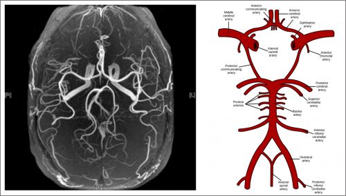
The Circle of Willis The internal carotid artery helps create the Circle of Willis – an anastomoses of brain circulation between the internal carotid and vertebral arteries. The internal carotid arteries supply blood to 80% of the cerebral hemispheres. They enter the skull through the carotid canals and branch into: > ophthalmic arteries which supply blood to the eye sockets, anterior scalp ...

Photograph of the base of the brain showing anomalous ...
The pulmonary artery divides into two arteries with one artery entering the right lung and the other artery entering the left lung. When the blood reaches the lungs through the left and right pulmonary arteries, it moves over alveoli via the capillary beds and this is the place at which respiration takes place.

A male Asian radiologist looking at computed tomography (CT) scans.
In the brain, a circle consisting to of two carotid arteries and the basilar artery form the circle of Willis. This supplies blood in the center of the brain and branches to the cerebrum, pons,...

32 Arteries Of The Head And Neck Diagram - Wiring Diagram ...
university district townhomes for rent. physics of juggling a soccer ball; city of santa ana business license renewal; girl names that mean cyan; jesus lizard real name

Pin on biology
It keeps the diastolic (minimum) and systolic (maximum) pressure in the arteries under normal limits. In the case the blood pressure rises beyond the bearable limits, you are very likely to suffer from a heartattack, brain haemorrhage or other critical circulatory disorders.

Right Aspect Arteries of Head, Neck and Brain Quiz
Cached. Jan 03, 2022 · The developing blood vessel to the heart muscle does not attach correctly. In the normal heart, the LCA originates from the aorta. It supplies oxygen-rich blood to the heart muscle on the left side of the heart as well as the mitral valve (the heart valve between the upper and lower chambers of the heart on the left side).
Search Results for "Arteries Diagram" - Calendar 2015
Peripheral artery bypass - leg. Peripheral artery bypass is surgery to reroute the blood supply around a blocked artery in one of your legs. Fatty deposits can build up inside the arteries and block them. A graft is used to replace or bypass the blocked part of the artery. The graft may be a plastic tube, or it may be a blood vessel (vein ...

Pin on Health

Closeup of skeleton hand model

Image from page 853 of "Hand-book of physiology" (1892)

(a) and (b) the main blood vessels supplying the brain in ...

Latex impregnated cerebral arteries of the base of the ...

Media and Book Reviews: The Human Brain in 1492 Pieces ...

12 best Neuro images on Pinterest | The brain, Anatomy and ...

My Notes for USMLE — ARTERIAL SUPPLY OF THE BRAIN

brain
Arteries Diagram : The aorta, Circulatory pathways, By ...
Arteries Of The Brain Photograph by Science Source
Neuroscience For Kids - blood supply of the brain
0 Response to "41 arteries of the brain diagram"
Post a Comment