39 acl and mcl diagram
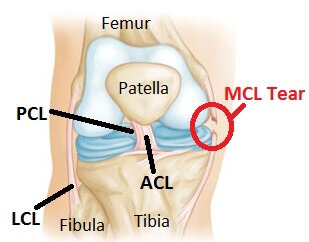
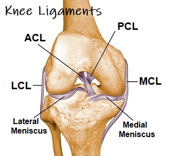
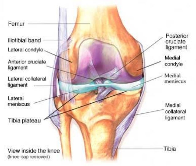
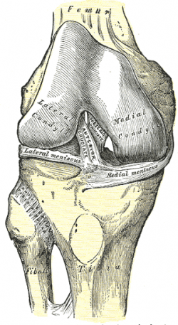
The medial collateral ligament (MCL) and lateral collateral ligament (LCL) are on the sides of the knee and prevent the joint from sliding sideways. The anterior cruciate ligament (ACL) and posterior cruciate ligament (PCL) form an "X" on the inside of the knee and prevent the knee from sliding back and forth. Diagrams showing the differences in intrinsic healing response of the anterior cruciate ligament (ACL; top) and medial collateral ligament (MCL; bottom), highlighting the lack of provisional scaffold (blood clot) formation within the ACL wound site as the key mechanism for ACL healing failure (reproduced with permission from Murray and Fleming 63).
Anterior cruciate ligament (ACL) ... (LCL and MCL) don't need surgery. However, when a cruciate ligament (ACL or PCL) is completely torn or stretched beyond its limits, the only option is ...

Acl and mcl diagram
An ACL tear often leads to the knee "giving out," and may require surgical repair. PCL (posterior cruciate ligament) strain or tear: PCL tears can cause pain, swelling, and knee instability. The Medial Collateral Ligament (MCL) is a two-part ligament found on the medial (inner) aspect of the knee. The deep part of this ligament attaches to the medial meniscus and the tibial joint line, and the superficial part starts and ends more superficially on the thigh and shin bone. The MCL acts to restrict excessive separation of the inside ... Unhappy Triad: Torn ACL, MCL and Meniscus An injury to a ligament in the knee often entails damage to other structures as well because of the complex construction of the joint. Surgeons refer to the so-called Unhappy Triad of torn ACL, MCL and meniscus— a triple-pronged injury that is often seen in knee patients.
Acl and mcl diagram. The medial collateral ligament (MCL) is a flat band of connective tissue that runs from the medial epicondyle of the femur to the medial condyle of the tibia and is one of four major ligaments that supports the knee. MCL injuries often occur in sports, being the most common ligamentous injury of the knee, and 60% of skiing knee injuries involve the MCL). Anterior to the intercondyloid eminence of the tibia, being blended with the anterior horn of the medial meniscus. The tibial attachment is in a fossa in front of and lateral to anterior spine, a rather wide area from 11 mm in width to 17 mm in AP direction.. For more detail on the anatomy of the ACL, please see this page: Anterior Cruciate Ligament (ACL) - Structure and Biomechanical Properties staged for optimal outcomes. Surgery uses an allograft or autograft to reconstruct the torn ACL and PCL ligaments, and may repair the MCL, LCL, and/or posterolateral corner of the knee if needed as well. Long-term complications after surgery include chronic pain, knee instability, arthrofibrosis, and loss of knee flexion ROM. ACL vs. MCL tears: Although symptoms of ACL and MCL tears are similar, a few key differences will help identify whether the injury affected the ACL or MCL. An ACL tear will have a more distinctive and loud popping sound than an MCL tear. The location of your pain and swelling could indicate either an ACL or MCL tear.
The medial collateral ligament (MCL) is one of four ligaments that is responsible for keeping the knee joint stable. (The other three are the anterior and posterior cruciate ligaments [ ACL and PCL] and the lateral collateral ligament [ LCL ].) The MCL connects the inner (medial) surfaces of the thigh bone (femur) and the shin bone (tibia) and ... Multiligament Knee Injury (ACL/PCL +/- MCL/PLC) Rehabilitation following surgery for multiligament knee injury (MLKI) or knee dislocation is an essential element of the treatment to achieve a full recovery. This protocol is intended to provide the user with instruction, direction, rehabilitative guidelines and functional goals. It is not meant ACL Tear and MCL Tear Treatment and Healing Time. For an ACL Tear: Treatment for an ACL tear will vary on two main factors: (a) The severity of the injury, and (b) the patient's lifestyle. For minor injuries or for a person who has either a sedentary lifestyle or low activity level, conservative treatment such as physical therapy may suffice. Acl And Mcl Diagram. Signs and symptoms of a medial collateral ligament (MCL) injury include The anterior cruciate ligament (ACL) and posterior cruciate ligament. The MCL is one of four major ligaments that supports the knee. either partial or complete ruptures in the ligament significantly increases the load on the ACL.
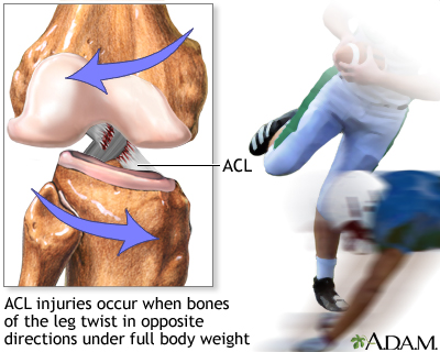
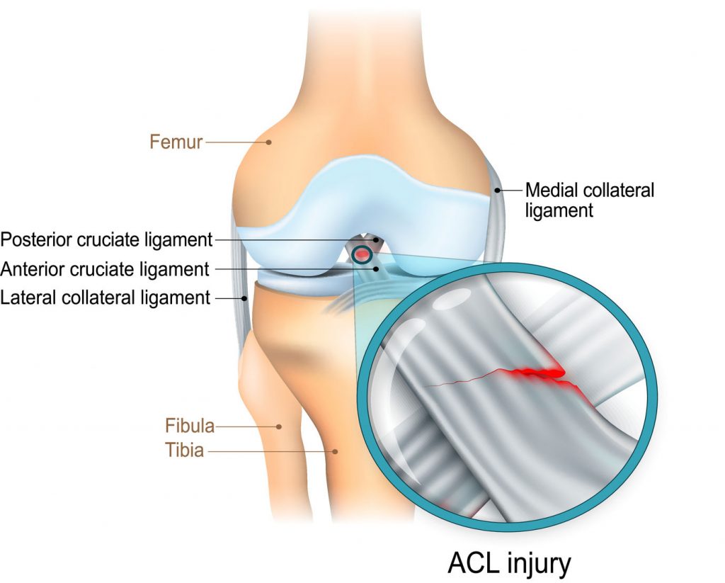
Here's a diagram of the knee that explains what and where the various ligaments are: Anatomy of the Knee. ACL = Anterior Cruciate Ligament (this has gone on me) MCL = Medial Collateral Ligmanet (also gone) - this is on the inside of my right knee. LCL = Lateral Collateral Ligament (also gone - outside of my right knee) Diagram of Knee Joint showing ACLAnterior Cruciate Ligament. The anterior cruciate ligament (ACL) is one of four critical ligaments that connects the bones of the knee joint. Its function is to provide stability to the knee and minimize the amount of stress that impacts the knee joint. The ACL accomplishes this by restraining the amount of ... Diagram of a torn ACL A torn ACL or ACL injury is a common occurrence among athletes and others who participate in sports. Females are 2 to 8 times more likely to experience an ACL injury than a male, depending on which sport is involved. Symptoms of a Torn MCL Diagram. Functions of the PCL and ACL. Mechanisms of Injury in the Knee. Diagram of a Torn PCL. First Healing Phase in a Knee Injury. Torn ACL Diagram. PLC and ACL Injury Treatment. Signs of a Torn PLC and ACL. ACL Tear Surgery Diagram. ACL, Medial Meniscus, and MLC Injury Diagram. Ligaments of the Knee Diagram.
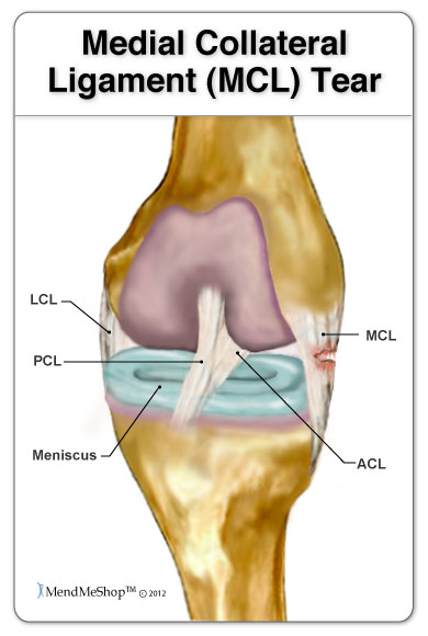
The exact location of MCL can be described as within the inner part of the knee from the bottom of the thighbone to a point on the top of shinbone, which is about 4-6 inches from the knee. The locations of ACL and MCL and where it can be torn are indicated in the following diagram: ACL vs. MCL Tears
MCL Tear Symptoms. Just as with an ACL tear, you may hear a popping sound when the injury occurs. Some of the most common symptoms of an MCL tear include: Pain on the inner side of the knee. Feeling the knee "give out". Swelling. The knee feels unstable. Pain when putting weight on the knee. Locked knee.
Unhappy Triad: Torn ACL, MCL and Meniscus An injury to a ligament in the knee often entails damage to other structures as well because of the complex construction of the joint. Surgeons refer to the so-called Unhappy Triad of torn ACL, MCL and meniscus— a triple-pronged injury that is often seen in knee patients.
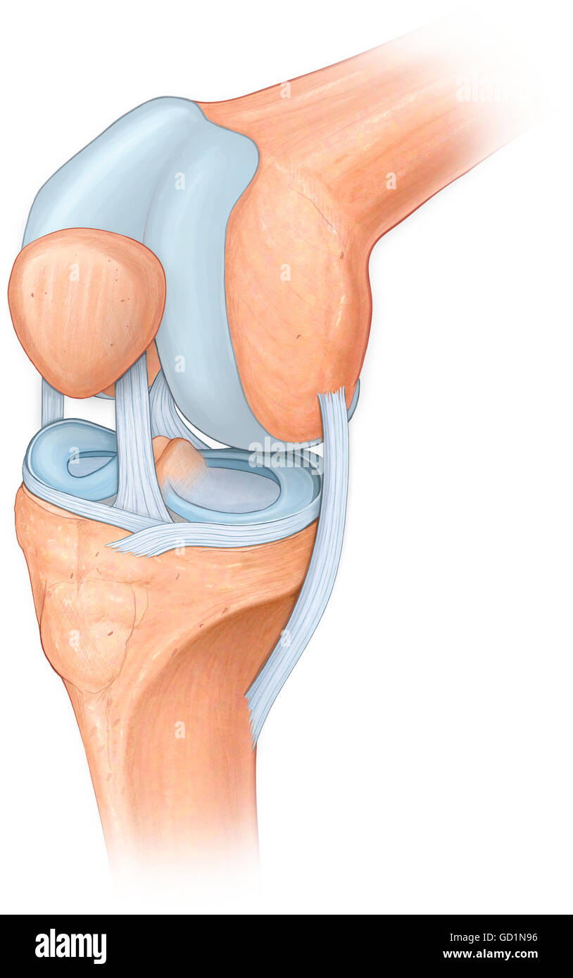
The Medial Collateral Ligament (MCL) is a two-part ligament found on the medial (inner) aspect of the knee. The deep part of this ligament attaches to the medial meniscus and the tibial joint line, and the superficial part starts and ends more superficially on the thigh and shin bone. The MCL acts to restrict excessive separation of the inside ...
An ACL tear often leads to the knee "giving out," and may require surgical repair. PCL (posterior cruciate ligament) strain or tear: PCL tears can cause pain, swelling, and knee instability.















:max_bytes(150000):strip_icc()/acl-surgery-rehab-exercises-3120748_final-2ceab43f9f984e6c84fbaf9d63046546.jpg)
0 Response to "39 acl and mcl diagram"
Post a Comment