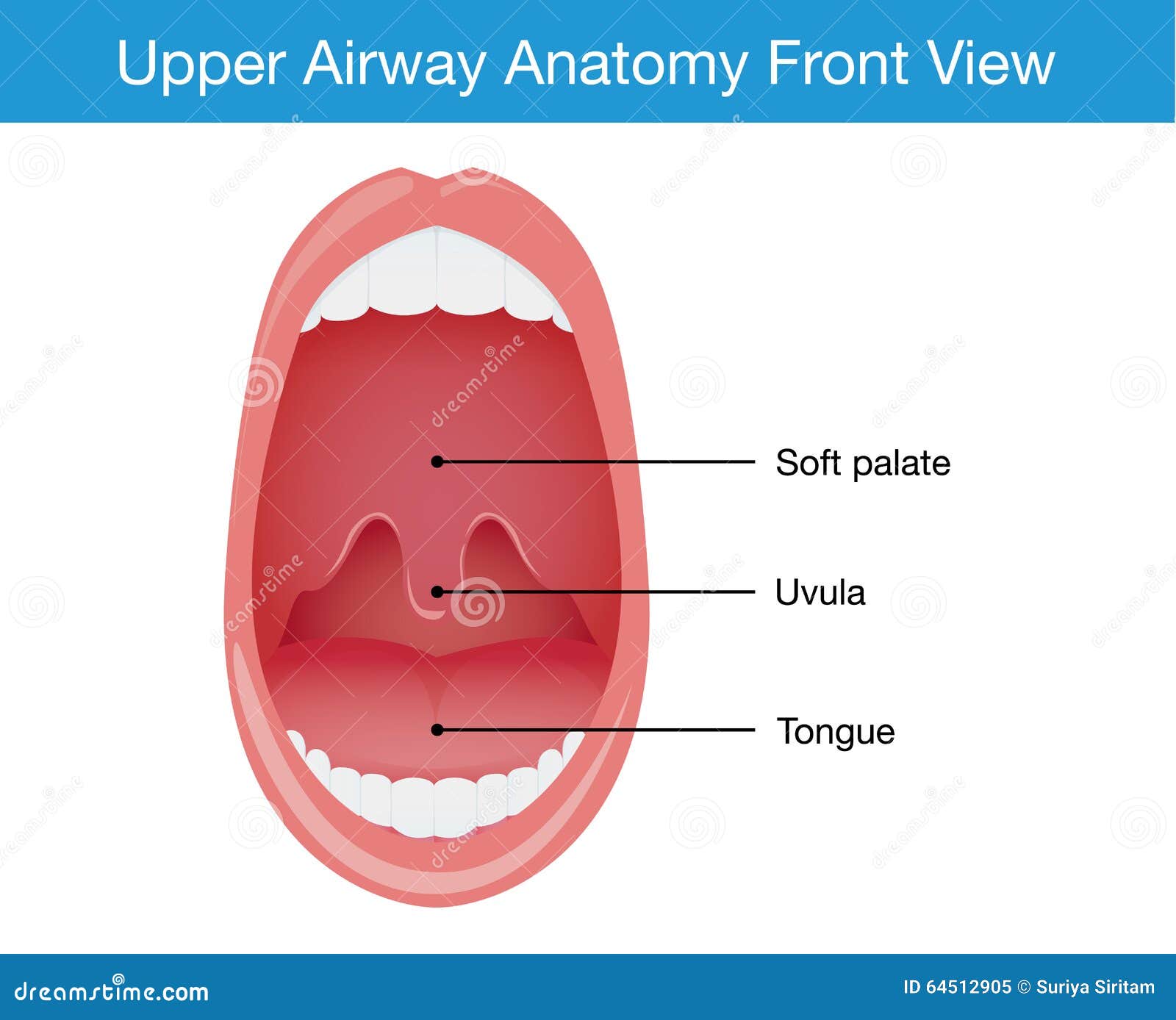
37 upper airway anatomy diagram
Start studying EMT Anatomy - Airway. Learn vocabulary, terms, and more with flashcards, games, and other study tools. Anatomy and physiology of the upper airway Proc Am Thorac Soc. 2011 Mar;8(1):31-9. doi: 10.1513/pats.201007-050RN. Authors Asli Sahin-Yilmaz 1 , Robert M Naclerio. Affiliation 1 Umraniye Education and Research Hospital, Department of Otolaryngology, Istanbul, Turkey. PMID: 21364219 DOI: 10.1513 ...
Structure and function of the upper airways.

Upper airway anatomy diagram
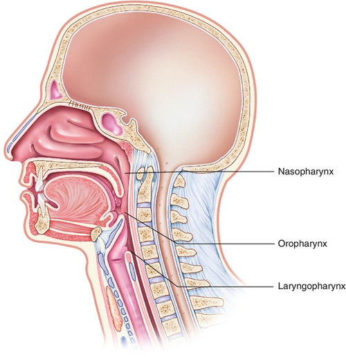
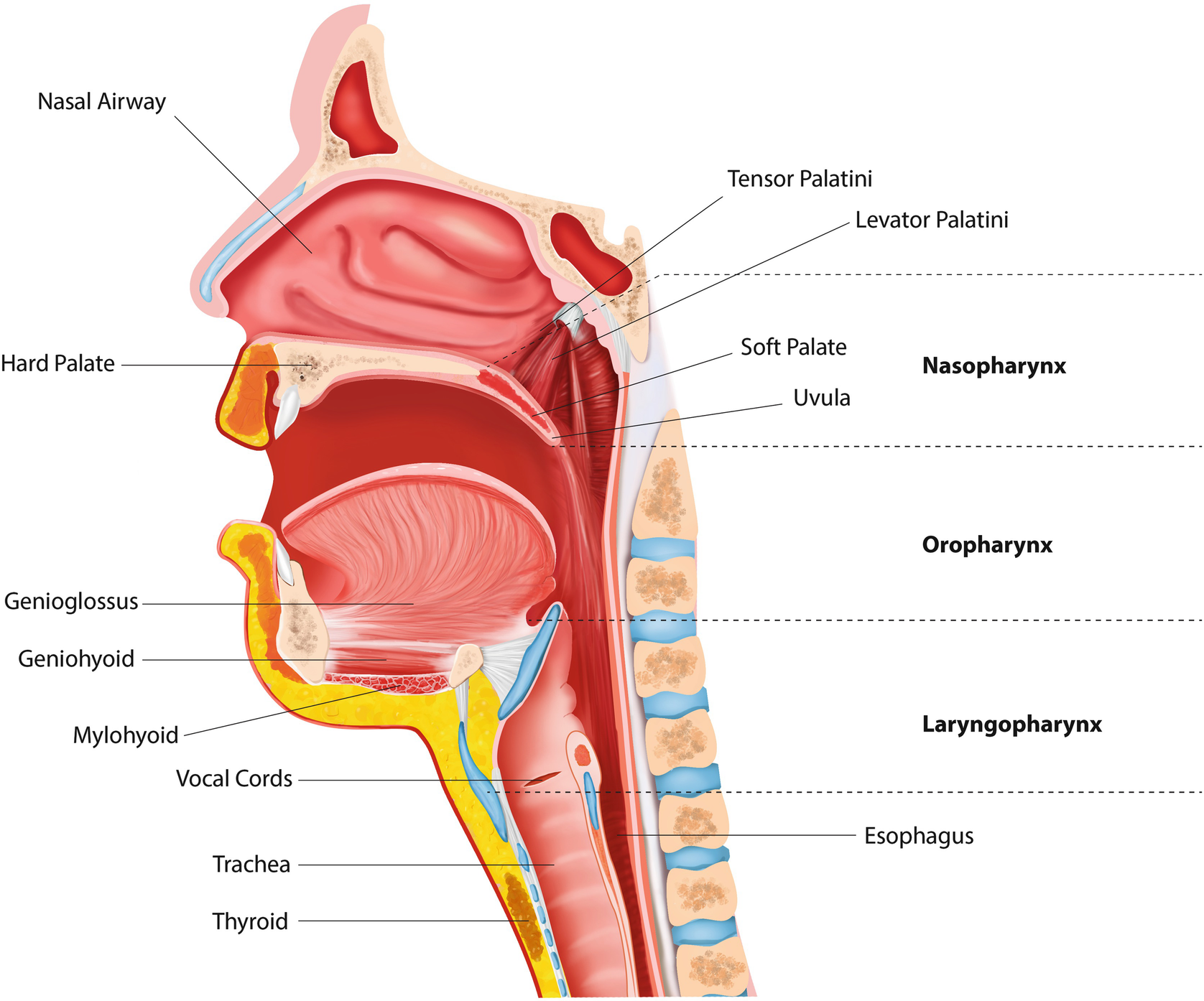
Anatomy and Physiology of the Upper Airway Asli Sahin-Yilmaz 1. x. Asli Sahin-Yilmaz. Search for articles by this author , and Robert M. Naclerio 2. x ... The complaint of alternating nasal obstruction with an acute upper respiratory tract infection shows how the nasal cycle becomes apparent during an illness . The upper airway consists of the pharynx and the nasal cavities; however, some authors include the larynx and trachea as well. The pharynx is can be divided into the nasopharynx, oropharynx, and laryngopharynx. The nose is composed of bone and cartilage, which are in turn attached to the facial skeleton. It is a pyramidal structure that is ... Basic Airway Anatomy Upper Airway. The upper airway is the "A" of the ABC's. As the entry point for oxygen any damage to, or blockage of, the structures in the upper airway can rapidly result in unconsciousness or death. The anatomy of the upper airway can be broken down into the nose, mouth, and throat.
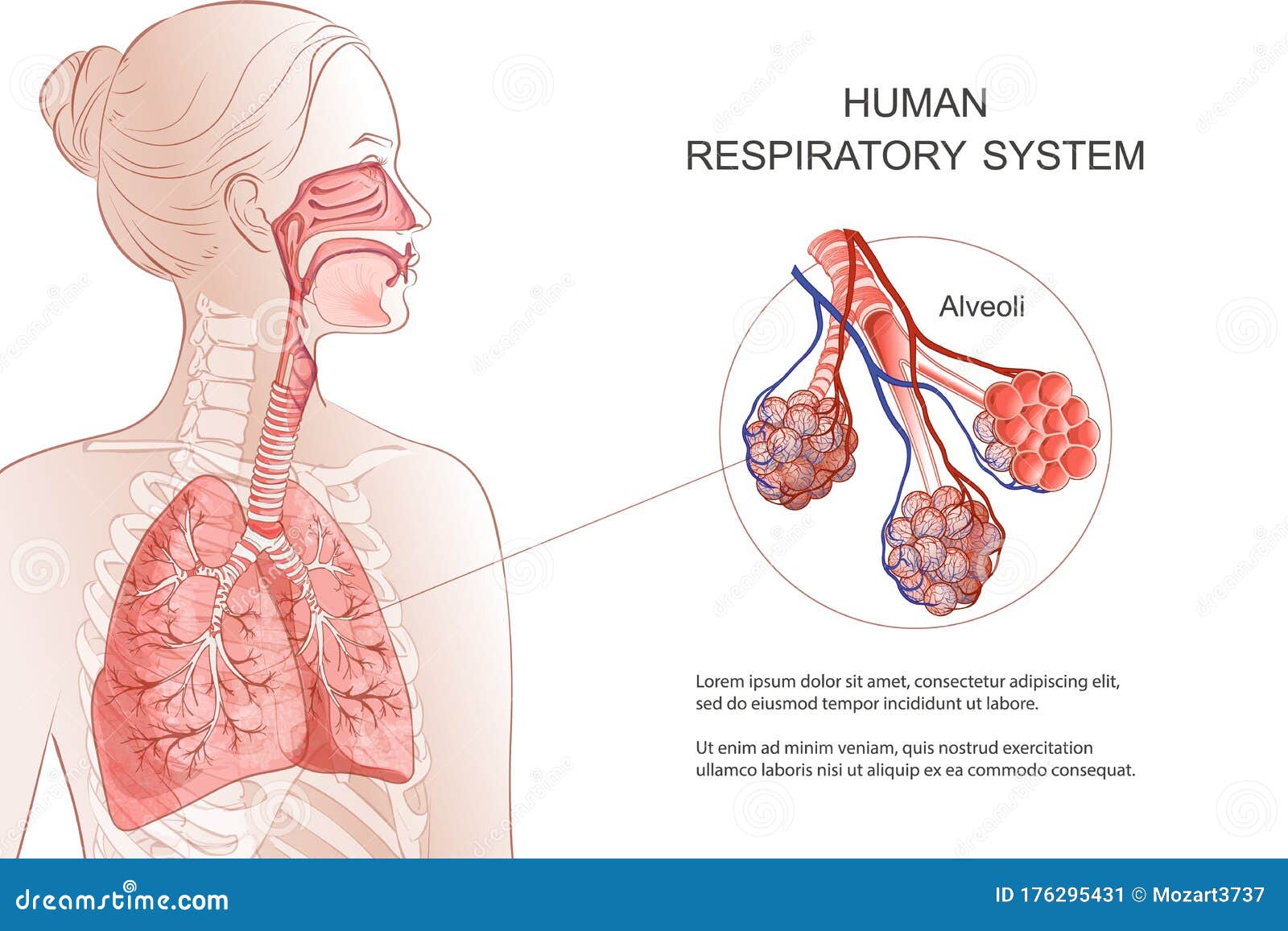
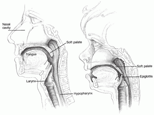
Upper airway anatomy diagram. Anatomy of infant and adult. Sagital section of the head and neck in (A) infant and (B) adult human. The food way and the airway are shaded in dark and light grey, respectively. (A) In infant human, the oral cavity is small, the tongue and palate … Anatomy and Physiology (A&P) of the Upper Airway. The airway begins at the tip of the nose and the lips and ends at the alveolocapillary membrane, through which gas exchange takes place between the air sacs of the lung (the alveoli) and the lung's capillary network. The airway consists of chambers and pipes, which conduct air with its 21% ... Week 7: Upper Airway Anatomy/ Airway anatomy. Pathway of air from nose to lungs. hyoid bone where is it found. thyroid cartilage. cricoid cartilage. Nose--> nasopharynx (occiput to soft palate)-->Oropharynx (sof…. level of C3. Pathway of air from nose to lungs. 15.3.2017 · Trachea Anatomy and Structure Tracheal Tissues and Membranes. Respiratory Mucosa: The innermost layer of the trachea, consisting of ciliated pseudostratified columnar epithelium and lamina propria (a thin layer of connective tissue), is covered with a sticky mucus coating produced by the goblet cells present in the region [1].
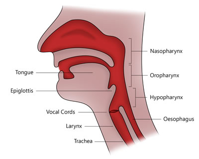
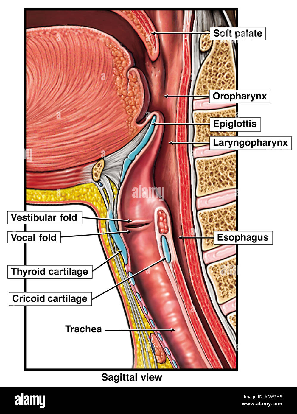
An understanding of these structures is important to the clinician involved in maintaining or reestablishing the normal airway. The fol … Anatomy and physiology of the upper airway Anesthesiol Clin North Am. 2002 Dec;20(4):733-45, v. doi: 10.1016/s0889-8537(02)00017-2. ... The airway is a continuous passageway but can be further categorized into the upper and lower airway. The upper airway begins from the nares and oropharynx to the vocal cords (Fig. 1a and b).The lower airway is located below the level of the vocal cords and extends distally. Upper Airway Anatomy Pediatric vs Adult Upper Airway Larger tongue in comparison to size of mouth Floppy epiglottis Delicate teeth, gums More superior larynx Funnel shaped larynx due to undeveloped cricoid cartilage Narrowest point at cricoid ring before ~8 years old. Upper Airway Anatomy. 28.11.2016 · What is the Larynx. The larynx, commonly called the voice box, is a 2-inch long cartilaginous tube connecting the back of the nose and the windpipe with each other.It is one of the most important structures of the respiratory system, also playing a crucial role in the production of speech in humans [1].. Where is the Larynx (Voice Box) Located
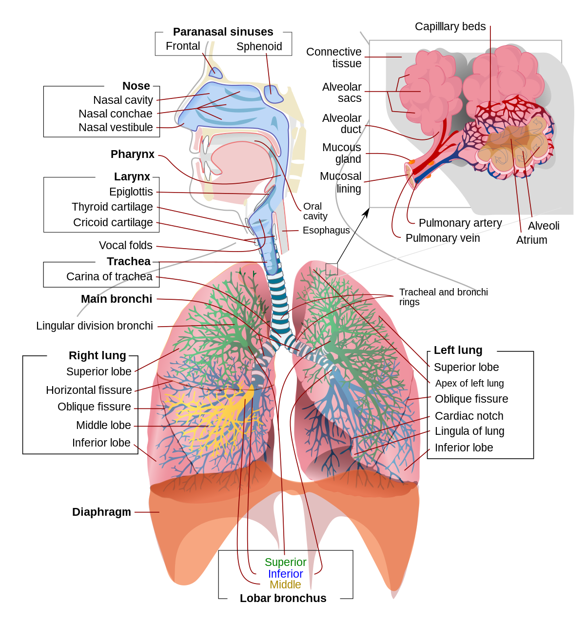
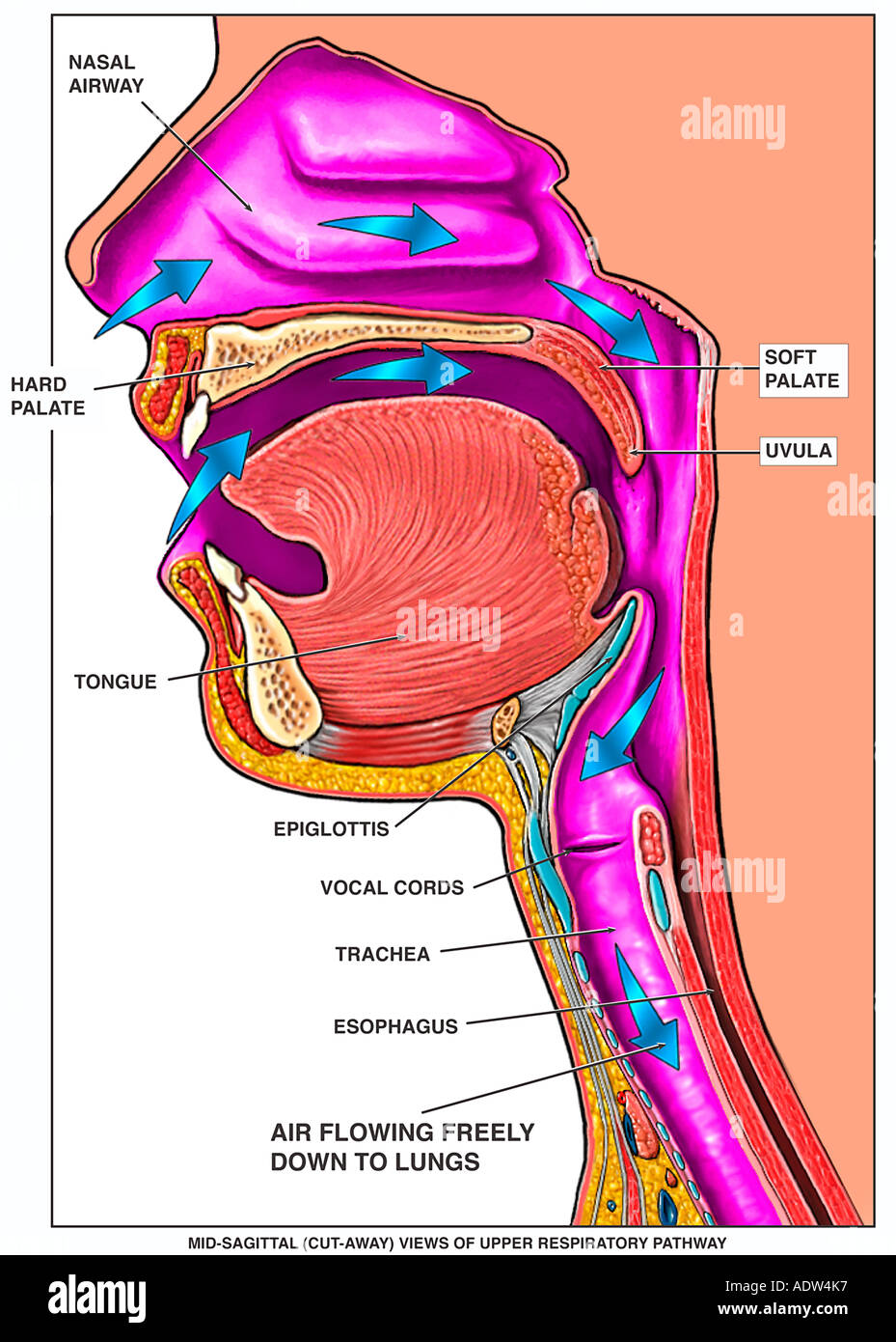
The upper respiratory system, or upper respiratory tract, consists of the nose and nasal cavity, the pharynx, and the larynx. These structures allow us to breathe and speak. They warm and clean the air we inhale: mucous membranes lining upper respiratory structures trap some foreign particles, including smoke and other pollutants, before the ... Upper Respiratory Tract Anatomy Nose Throat Mouth. Click Images to Large View Upper Respiratory Tract Anatomy Nose Throat Mouth. Intubation. Equine. Normal. Upper Airway Labeled. Oral Airway Anatomy. Bronchoscopy. Upper vs Lower Airway. Airway Anatomy. Airway anatomy can be divided into the upper airway and the lower airway. First responders need to be familiar with respiratory system anatomy in order to keep patients healthy, breathing and adequately ventilated. Below are detailed graphics of both the upper and lower respiratory tracts. Upper Airway Anatomy Posted on June 7, 2016 by admin. Picture Of Female Reproductive System Diagram 1024×1204 Diagram - Picture Of Female Reproductive System Diagram 1024×1204 Chart - Human anatomy diagrams and charts explained. This diagram depicts Picture Of Female Reproductive System Diagram 1024×1204 with parts and labels.
IT_AM 1.1 Describe the basic structural anatomy of the upper airway including the larynx BT_AM 1.1 Describe the anatomy of the upper airway, larynx and trachea …. To get the most benefit out of this post, print out the diagram below. Some of the questions need you to draw on it. Name structure A (be specific!)
This diagram outlines the main anatomical structures of the upper airway that are important when inserting a tracheostomy tube. Anatomy of the upper airway The tracheostomy is the procedure of creating an artificial opening into the front of the trachea, to bypass the airway superior to this where there may be a cancer, obstruction, or to ...

Airway anatomy 1. Anatomy of theAnatomy of the Upper AirwayUpper Airway Dr. Saurabh Barde, Anaest hesiology, GMCH, Nagpur. 2. Dr. Saurabh Barde, Anaest hesiology, GMCH, Nagpur. The Upper Airway is a Continuation of the Respiratory System 3.

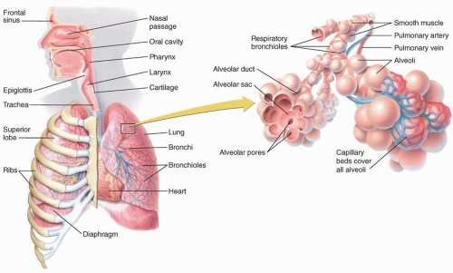
The upper respiratory tract is made up of the: Nose. Nasal cavity. Sinuses. Larynx. Trachea. The lower respiratory tract is made up of the: Lungs. Bronchi and bronchioles. Air sacs (alveoli) Lungs. The lungs take in oxygen. Your body's cells need oxygen to live and carry out their normal functions.

Upper respiratory tract: this part of the respiratory system extends from the nose to the trachea; sinuses ring the nose and upper jaw [NHLBI 2017.] Upper Respiratory Tract Diagram Frontal sinus Nostril Oral cavity Trachea. Title: B1162-002972-16_Sanofi_CSO_Allegra_Module1_Diagram_ADL02.indd

Upper Respiratory System. Create healthcare diagrams like this example called Upper Respiratory System in minutes with SmartDraw. SmartDraw includes 1000s of professional healthcare and anatomy chart templates that you can modify and make your own. 21/22 EXAMPLES. EDIT THIS EXAMPLE.

Upper Respiratory Tract Structural and Functional Anatomy Nose and Nasal Cavity. The nostrils, the two round or oval holes below the external nose, are the primary entrance into the human respiratory system [5].Lying just after the nostrils are the two nasal cavities, lined with mucous membrane, and tiny hair-like projections called cilia [6].During inhalation, the air passes into the nasal ...
9.11.2021 · Upper respiratory tract. The upper respiratory tract refers to the parts of the respiratory system that lie outside the thorax, more specifically above the cricoid cartilage and vocal cords.It includes the nasal cavity, paranasal sinuses, pharynx and the superior portion of the larynx.Most of the upper respiratory tract is lined with the pseudostratified ciliated columnar …

Objectives Review anatomy relevant to airway managementReview anatomy relevant to airway management Relate key differences in airway structures and how they influence successful bag mask ventilation (BMV) Describe the process of opening the airway and maitii itintaining it Describe the indications, limitations, proper sizing and contraindications of BLS airwaysizing, and contraindications of ...
The airway, or respiratory tract, describes the organs of the respiratory tract that allow airflow during ventilation. [1][2][3]They reach from the nares and buccal opening to the blind end of the alveolar sacs. They are subdivided into different regions with various organs and tissues to perform specific functions. The airway can be subdivided into the upper and lower airway, each of which ...

21.1.2018 · The nose is the body's primary organ of smell and also functions as part of the body's respiratory system. Air comes into the body through the nose. As …

The upper airway system comprises the nose and the paranasal cavities (or sinuses), the pharynx (or throat), and partly also the oral cavity, since it may be used for breathing.The lower airway system consists of the larynx, the trachea, the stem bronchi, and all the airways ramifying intensively within the lungs, such as the intrapulmonary bronchi, the bronchioles, and the …

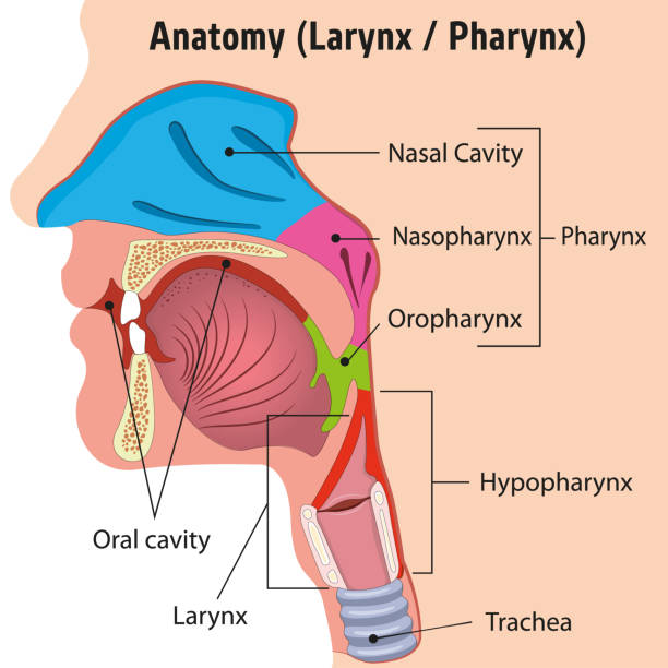
The pharynx is a musculomembranous tube that functions as both part of the alimentary canal and an airway of the respiratory system. It is divided into three parts: the nasopharynx, oropharynx, and laryngopharynx. Image from Human Anatomy Atlas. Nasopharynx. The nasopharynx is the portion of the pharynx that begins at the rear of the nasal cavity.
Respiratory. The respiratory system, which includes air passages, pulmonary vessels, the lungs, and breathing muscles, aids the body in the exchange of gases between the air and blood, and between ...
Our new CrystalGraphics Chart and Diagram Slides for PowerPoint is a collection of over 1000 impressively designed data-driven chart and editable diagram s guaranteed to impress any audience. They are all artistically enhanced with visually stunning color, shadow and lighting effects. ... "Anatomy of the Upper Airway" is the property of its ...
23.11.2020 · The Human Respiratory System - explore anatomy of the upper and lower respiratory tracts, from nasal passages to the lungs, using interactive diagrams.

18.11.2021 · The main function of the trachea is to transport air from the upper respiratory tract to the lungs. Air that enters the trachea is warmed and moisturized before moving on to the lungs. Mucus on the trachea walls can catch debris or particles. This debris is then transported upward by cilia, tiny hair-like structures that remove it from the airway.

The upper airway is full of muscles, the anatomy of which is the nightmarish province of ENT and maxillofacial surgeons. For the intensivist, it will likely suffice to know that their function in respiration is basically to not collapse. And to collapse, apparently, is their natural tendency.

Upper airway impairment manifested in combination with breathing pattern disorders (e.g., vocal cord dysfunction) Functional DB (a subdivision of thoracic and extrathoracic DB) No structural or functional alterations directly associated with the symptoms of DB (e.g., phrenic nerve palsy, myopathy, and diaphragmatic eventration ((one leaf of diaphragm elevated …

Basic Airway Anatomy Upper Airway. The upper airway is the "A" of the ABC's. As the entry point for oxygen any damage to, or blockage of, the structures in the upper airway can rapidly result in unconsciousness or death. The anatomy of the upper airway can be broken down into the nose, mouth, and throat.

The upper airway consists of the pharynx and the nasal cavities; however, some authors include the larynx and trachea as well. The pharynx is can be divided into the nasopharynx, oropharynx, and laryngopharynx. The nose is composed of bone and cartilage, which are in turn attached to the facial skeleton. It is a pyramidal structure that is ...

Anatomy and Physiology of the Upper Airway Asli Sahin-Yilmaz 1. x. Asli Sahin-Yilmaz. Search for articles by this author , and Robert M. Naclerio 2. x ... The complaint of alternating nasal obstruction with an acute upper respiratory tract infection shows how the nasal cycle becomes apparent during an illness .


0 Response to "37 upper airway anatomy diagram"
Post a Comment